
Figures & data
Figure 1 Pathological changes in a rheumatoid arthritis joint. In established RA, the inflamed synovial membrane forms a pannus, due to infiltration of peripheral blood cells and proliferation of fibroblast-like synoviocytes. These cells are highly activated releasing pro-inflammatory mediators and autoantibodies within the joint sustaining the inflammatory process. This is accompanied by cartilage damage and osteoclast-mediated bone erosion leading to invasion of the pannus tissue and irreversible deformation of the joint.
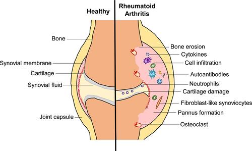
Figure 2 Two signal model for classical NLRP3 inflammasome activation by TLR4. During the priming stage, activation of TLR4 by MAMPs or DAMPs upregulates NLRP3 and pro-IL-1β expression through NF-κB activation. This is closely followed by activation and assembly of the NLRP3 inflammasome, which can be induced by various stimuli including K+ efflux, Ca2+ signaling, mitochondrial dysfunction, and lysosomal rupture. Upon activation, caspase-1 cleaves pro-IL-1β and GSDMD resulting in pyroptosis and IL-1β release. Created with BioRender.com.
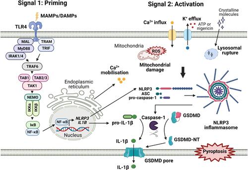
Table 1 Endogenous Toll-Like Receptor Ligands Associated with Rheumatoid Arthritis
Table 2 Summary of Some of the Main Associations of TLRs with RA Pathogenesis in Human and Animal Disease Models