Figures & data
Figure 1 Fibrotic Nonspecific Interstitial Pneumonia in a patient with a longstanding Mixed Connective Tissue Disease. The pattern is characterized by diffuse Ground Glass Opacities (yellow arrow), subpleural sparing (blue arrow), and presence of traction bronchiectasis (red arrow).
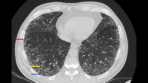
Table 1 Proposed Classification Criteria for Mixed Connective Tissue Disease
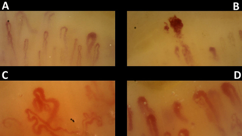
Figure 2 Nailfold Videocapillaroscopy in a healthy subject (A) compared with a patient with Mixed Connective Tissue Disease (B–D). The pattern is characterized by the presence of multiple Giant Capillaries and neoangiogenesis.