Figures & data
Figure 1 Colonoscopy showed ileocecal mass (A). Histopathological examination showed moderately and poorly differentiated adenocarcinoma (B).
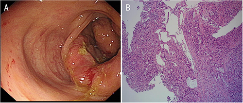
Figure 2 CT showed lymph node enlargement in the left supraclavicular fossa (A), thickening of the right colon wall with multiple metastatic lymph nodes in the abdomen (C and E). After conversion treatment, the enlarged lymph nodes (B and D) and primary tumor (F) were significantly reduced.
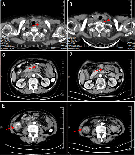
Figure 3 Specimen examination showed a 4cm×1.5cm bulged mass located ileocecal valve with hard gray texture on section (A). Histological analysis of the specimen and resected 14 lymph nodes revealed no malignancy (B).
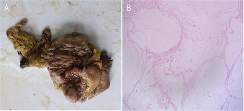
Figure 4 Timeline.
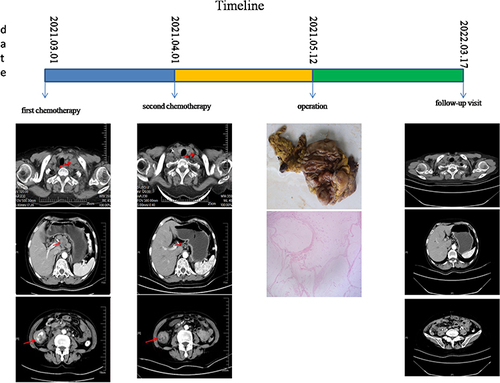
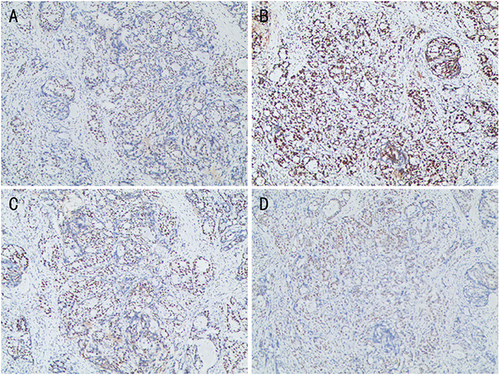
Figure 5 Immunohistochemistry showed MLH (+) (A), MSH2 (+) (B), MSH6 (+) (C), PMS2 (+) (D). That is mismatch-repair-proficient (pMMR) and microsatellite stable (MSS).