Figures & data
Table 1 EpitopesTable Footnotea,Table Footnoteb of Trichomonas vaginalis fructose-1,6-bisphosphate aldolase are reactive with MAbs and human sera
Table 3 EpitopesTable Footnotea of Trichomonas vaginalis glyceraldehyde-3-phosphate dehydrogenase are reactive with MAbs and human sera
Table 4 List of synthetic 15-mer peptides of representative epitopes of ALD, ENO, GAP, and ACT that are reactive with positive control women and men sera
Figure 1 Sequence analyses of Trichomonas vaginalis fructose-1,6-bisphosphate aldolase.
Abbreviations: ALD, aldolase; Ca, Candida albicans; Ec, Escherichia coli; Hs, Homo sapiens; M, men; MAbs, monoclonal antibodies; Ng, Neisseria gonorrhoeae; Sa, Staphylococcus aureus; Sc, Saccharomyces cerevisiae; Spn, S. pneumoniae; Spy, Streptococcus pyogenes; Tp, Treponema pallidum; W, women; ALD12 and ALD64, designate MAbs to aldolase as per .
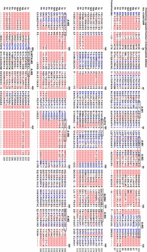
Figure 2 Hydrophobicity and antigenicity profiles of Trichomonas vaginalis α-enolase, aldolase, and glyceraldehyde-3-phosphate dehydrogenase proteins.
Abbreviations: A and ALD, fructose-1,6-bisphosphate aldolase; E and ENO, α-enolase; G and GAP, glyceraldehyde-3-phosphate dehydrogenase; M, men; MAb, monoclonal antibody; W, women; ALD13, ALD55, ALD11, ALD25, and B44, designate MAbs to alpha-enolase, ALD12/64 designates a MAb to aldolase, and ALD32, ALD30, and B43 designate MAbs to glyceraldehyde-3-phosphate dehydrogenase.
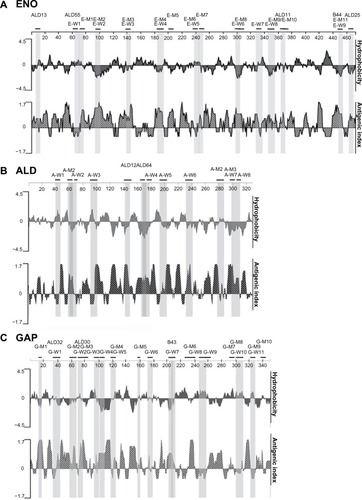
Figure 3 Representative experiment showing dot blots of individual 15-mer peptide epitopes.
Abbreviations: ACT, α-actinin; ALD, fructose-1,6-bisphosphate aldolase; ENO, α-enolase; GAP, glyceraldehyde-3-phosphate dehydrogenase.
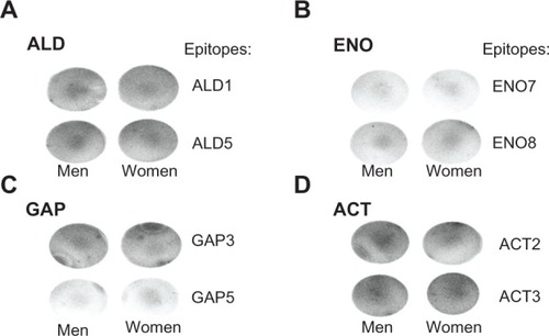
Figure 4 Representative experiments in duplicate dot blots of combinations of 15-mer peptide epitopes (A) and densitometric scans of reactive dot blots (B).
Abbreviations: ACT, α-actinin; ALD, fructose-1,6-bisphosphate aldolase; con, control; ELISA, enzyme-linked immunosorbent assay; ENO, α-enolase; GAP, glyceraldehyde-3-phosphate dehydrogenase glyceraldehyde; neg, negative.
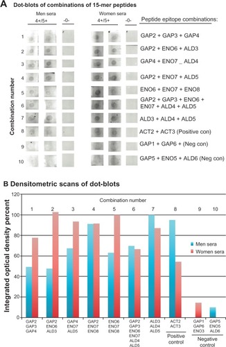
Figure 5 The 111 amino acid sequence and expression and purification of the rSOE encoding epitopes of GAP, ENO, and ALD.
Abbreviations: ALD, fructose-1,6-bisphosphate aldolase; ENO, α-enolase; GAP, glyceraldehyde-3-phosphate dehydrogenase; His6, hexahistidine; MAb, monoclonal antibody; mw, molecular weight; Ni2+, nickel; NTA, nitrilotriacetic acid; rSOE, recombinant protein of sequences of epitopes; SDS-PAGE, sodium dodecyl sulfate polyacrylamide gel electrophoresis; SOE, sequences of epitopes.
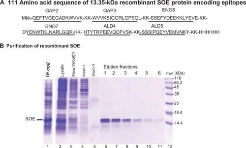
Figure 6 Immunodetection of the rSOE by ELISA (A), dot blot (B), and immunoblotting after SDS-PAGE (C).
Abbreviations: ELISA, enzyme-linked immunosorbent assay; His6, hexahistidine; MAb, monoclonal antibody; mw, molecular weight; rSOE, recombinant protein of sequences of epitopes; SDS-PAGE, sodium dodecyl sulfate polyacrylamide gel electrophoresis.
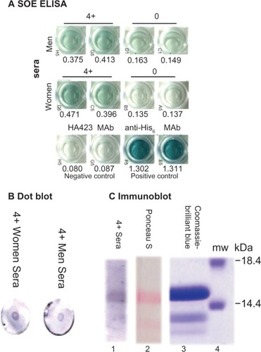
Table 2 EpitopesTable Footnotea of Trichomonas vaginalis α-enolase are reactive with MAbs and human sera