Figures & data
Table 1 Severity index for the dermatitis score
Table 2 Primers used in quantitative real-time polymerase chain reaction
Figure 1 Schematic representation of the airway lumen and surrounding smooth muscle.
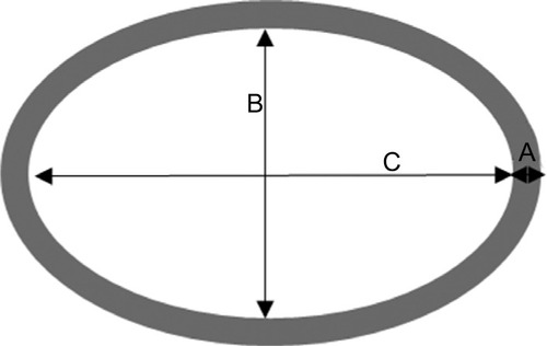
Table 3 Scoring system for measuring the amount of mucus plug in the airway
Figure 2 Atopic dermatitis-like skin inflammation, pruritus, and amplification of Th2 inflammatory reactions.
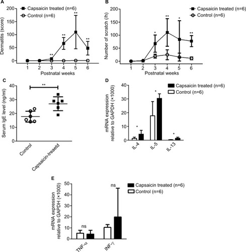
Figure 3 Eosinophil infiltration and elevated expression of Th2 cytokines.
Abbreviations: BAL, bronchoalveolar lavage; ns, non significant.
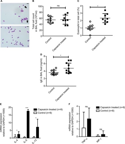
Figure 4 Airway remodeling and airway hyperresponsiveness.
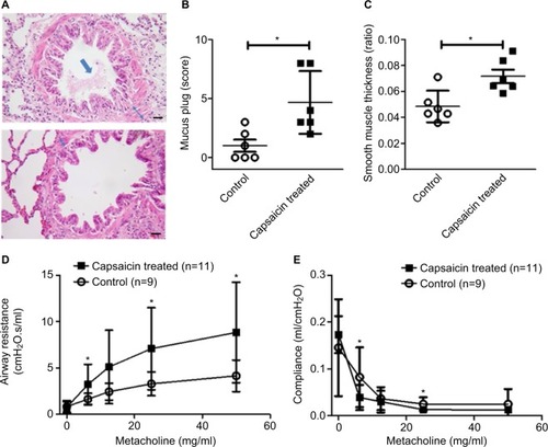
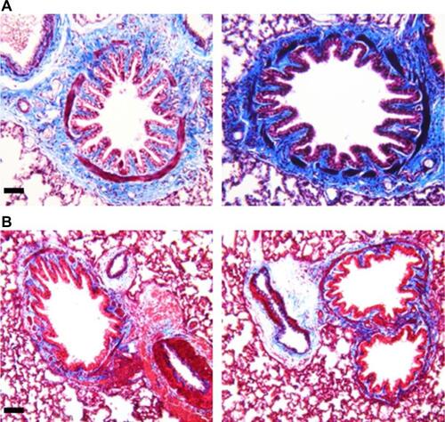
Figure S1 Increased collagen deposition in the airways.
Note: Representative slides revealed relative abundance of collagen deposition in the airways from the capsaicin-treated rats (A) compared to control rats (B) (scale bar=50 μm).