Figures & data
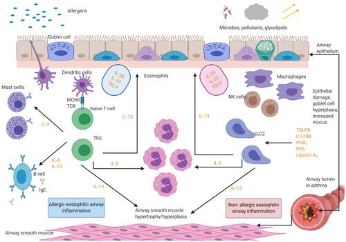
Figure 1 Eosinophilic asthma inflammatory pathways. There are two aetiologies for eosinophilic inflammation in asthma: an allergic pathway triggered by allergens and a non-allergic mechanism triggered by microbes, pollutants and glycolipids. The key mediators in the pathways are depicted below. Eosinophils release cationic proteins,Citation57 which lead to bronchial epithelial tissue damage, thus causing airways hyper-responsiveness. Eosinophils also lead to airway smooth muscle cell proliferation through increased eosinophils adhesion caused by the release of cationic proteins and the eosinophilic effect on transforming growth factor-β1 and gene coding of wingless/integrase-1 signaling. IL-13 triggers mucus hyper-secretion. Figure created with BioRender.com.
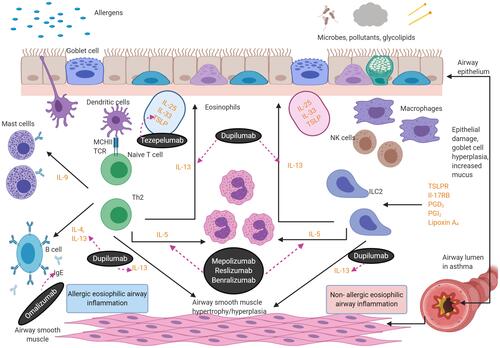
Table 1 Licensed Biologics Used for Severe Asthma
Figure 2 Licensed mediator cascade of biologics used in eosinophilic asthma. The figure below shows the targets for the licensed biologics. Omalizumab is an IgE-blocker. Mepolizumab, reslizumab and benralizumab are anti IL-5 agents. Dupilumab is an Anti-IL-4/anti-IL-13 agent. Figure created with BioRender.com.