Figures & data
Figure 1 The expression level of miR-21-5p was increased in rats with OVA-induced asthma. Histological changes were analyzed by H&E staining assay (A) (Scale bar = 50 μm) and Masson staining assay (B) (Scale bar = 50 μm). The levels of epithelial cell adhesion factor and stromal cell markers were analyzed by Western blot (C and D, n=6). The expression level of miR-21-5p were analyzed by qRT-PCR analysis (E, n=6) and FISH (F). (G) Representative immunofluorescent staining of lung tissues from control and asthma group showing the expression of α-SMA and E-cadherin. Data were expressed as mean ± SD. *Indicates P < 0.05.
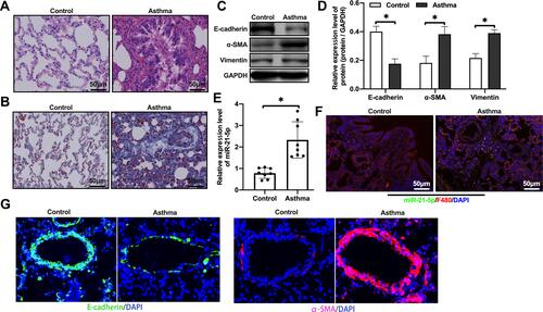
Figure 2 Alveolar macrophages stimulated by LPS could secreted exosomes with high levels of miR-21-5p. The expression level of miR-21-5p in macrophages were analyzed by qRT-PCR analysis (A, n=6). Exosomes were isolated from the cell culture supernatant of macrophages by high-speed centrifugation, and their morphology was verified by transmission electron microscopy (B) and nanoparticle tracking analysis of the particle size distribution (C). The expression of exosomal surface markers were analyzed by Western blot (D, n=5). The expression level of miR-21-5p in the exosomes were analyzed by qRT-PCR analysis (E and F). The concentration of exosomes (vesicles/mL) was measured by nanoparticle tracking analysis (n=6). Data were expressed as mean ± SD. *Indicates P < 0.05.
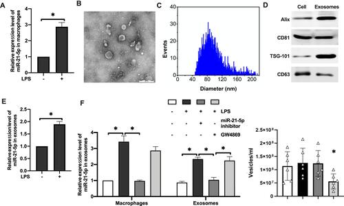
Figure 3 MiR-21-5p was transported to tracheal epithelial cells through exosomes and promoted EMT. The exosomes were labeled with fluorescent dye PKH67 and observed under a fluorescence microscope (A). The level of miR-21-5p in rat tracheal epithelial cells cocultured with exosomes isolated from the same amount of culture medium were analyzed by qRT-PCR analysis (B and F) (n=3–6). The levels of epithelial cell adhesion factor and stromal cell markers were analyzed by Western blot (C, D and G) (n=5). The levels of α-SMA and vimentin were analyzed by Immunofluorescence assay (E). Data were expressed as mean ± SD. *Indicates P < 0.05.
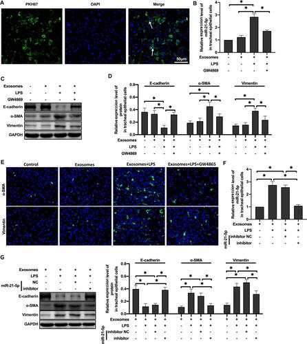
Figure 4 MiR-21-5p in exosomes derived from LPS-treated alveolar macrophages targeted Smad7 in rat tracheal epithelial cells. Luciferase was determined using plasmids containing wild-type or mutant 3ʹUTR Smad7 proteins and the binding site of miR-21- 5p (A–C) (n=3–6). The expression of Smad7 and TGFβ1 protein were analyzed by Western blot (D and E) (n=3). Data were expressed as mean ± SD. *Indicates P < 0.05.
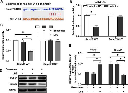
Figure 5 MiR-21-5p promoted EMT of rat airway epithelial cells by inhibiting the expression of Smad7. The expression of TGFβ1, TGFβRII and Smad7 protein were analyzed by Western blot (A and B) (n=3–4). The expression of TGFβ1, TGFβRII and Smad7 mRNA were analyzed by qRT-PCR analysis (C) (n=3–4). The level of miR-21-5p in rat tracheal epithelial cells were analyzed by qRT-PCR analysis (D) (n=3–5). The expression of Smad7, TGFβ1, TGFβRII, p-Smad2 and p-Smad3 protein were analyzed by Western blot (E and F) (n=3). The levels of epithelial cell adhesion factor and stromal cell markers were analyzed by Western blot (G and H) (n=3). Data were expressed as mean ± SD. *Indicates P < 0.05.
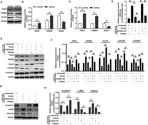
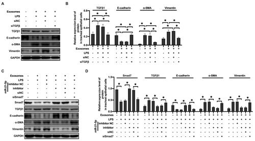
Figure 6 MiR-21-5p in exosomes produced by LPS-treated macrophages promoted EMT of rat tracheal epithelial cells through TGFβ1/Smad signal pathway. The levels of epithelial cell adhesion factor and stromal cell markers and TGFβ1 were analyzed by Western blot (A and B) (n=3–4). The levels of epithelial cell adhesion factor and stromal cell markers, Smad7 and TGFβ1 were analyzed by Western blot (C and D) (n=3–4). Data were expressed as mean ± SD. *Indicates P < 0.05.