Figures & data
Figure 1 The expression of circERBB2 was upregulated in the biopsy samples of patients with asthma and PDGF-BB-stimulated ASMCs. (A) QRT-PCR quantification of circERBB2 level in biopsy samples collected from patients with asthma (n=45) and healthy donors (n=45). (B-D) ASMCs were stimulated with PDGF-BB (25 ng/mL) for 24 hours, the levels of circERBB2, IGF1R, and miR-98-5p were detected by qRT-PCR assay. *p < 0.05, **p < 0.01.
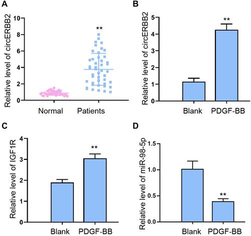
Figure 2 CircERBB2 knockdown suppressed PDGF-BB -stimulated proliferation and migration of ASMCs. (A) Expression of circERBB2 in ASMCs transfected with shcircERBB2-1, shcircERBB2-2, or negative control (shNC) were detected by qRT-PCR experiment. (B, C) ASMCs were treated with PDGF-BB and depleted of circERBB2. Cell proliferation was detected by CCK-8 assay. (D) Cell migration was detected by Transwell assay. (E) The expression of PCNA and MMP-9 were measured by Western blotting. (F) Apoptosis of ASMCs was measured by flow cytometry. (G) The expression of Bax, cleaved caspase-3, and Bcl-2 was measured by Western blot analysis. (H, K) The secreted levels of IL-1β, IL-6, TNF-α, and IL-13 were determined by ELISA assay. Shcirc-1, shcircERBB2-1; shcirc-2, shcircERBB2-2; shNC, negative control. **p < 0.01.
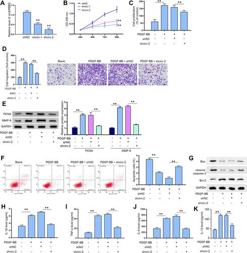
Figure 3 CircERBB2 directly interacted with miR-98-5p. (A) Predicted binding sites of miR-98-5p on circERBB2. (B) QRT-PCR assay to detect the transfection efficacy of miR-98-5p mimics in BC cells. (C) Luciferase activity of circERBB2-WT and circERBB2-MUT after transfection with miR-98-5p mimics or NC. (D, E) RNA pulldown (D) and RIP experiment (E) to evaluate the interaction between miR-98-5p and circERBB2. (F) QRT-PCR assay to measure the level of miR-98-5p in ASMCs after circERBB2 depletion. (G) QRT-PCR quantification of circERBB2 and miR-98-5p levels in biopsy samples collected from patients with asthma (n=45). **p < 0.01.
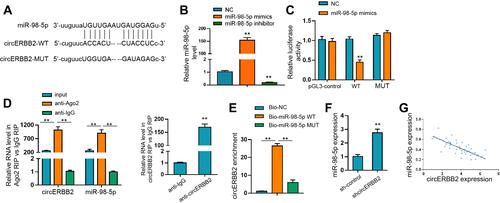
Figure 4 MiR-98-5p suppressed the PDGF-BB-stimulated proliferation and migration of ASMCs. (A) Viability of ASMCs after transfection with miR-98-5p mimics was determined by CCK-8. (B, C) CCK-8 assay and Transwell assay to determine viability and migration of ASMCs treated with PDGF-BB, shcircERBB2 and miR-98-5p inhibitors. Histogram showed the portion of viable or migrated cells. (D) Flow cytometry to detect cell migration. Histogram showed the portion of apoptotic cells. (E) The expression of Bax, cleaved caspase-3, and Bcl-2 was measured by Western blot analysis. **p < 0.01.
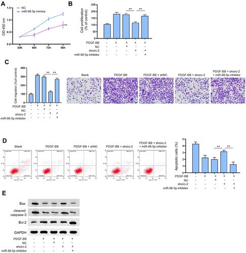
Figure 5 MiR-98-5p interacted with 3ʹUTR of IGF1R and IGF1R regulated PDGF-BB-stimulated proliferation and migration of ASMCs. (A) Predicted binding site of miR-98-5p on 3ʹUTR of IGF1R. (B) Luciferase activity of IGF1R-WT and IGF1R-MUT after transfection with miR-98-5p mimics or NC. (C) QRT-PCR assay to measure the level of IGF1R in ASMCs after miR-98-5p transfection. (D) QRT-PCR assay to measure the level of IGF1R after transfection with shIGF1R-1, shIGF1R-2 or shNC. (E, F) CCK-8 assay to determine cell viability. (G) Transwell assay to detect cell migration. The histogram showed the portion of migrated cells. (H) Western blotting assay to detect the expression of IGF1R. **p < 0.01.
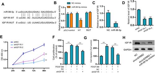
Figure 6 IGF1R mediated miR-98-5p-suppressed proliferation and migration of ASMCs. ASMCs were treated with PDGF-BB, and transfected with miR-98-5p, IGF1R overexpressing vector (IGF1R OE) or negative controls. Then the proliferation (A), migration (B), and apoptosis (C) of ASMCs were determined by CCK-8, Transwell, and flow cytometry assay, respectively. (D) The expression of Bax, cleaved caspase-3, and Bcl-2 was measured by Western blot analysis. **p < 0.01.
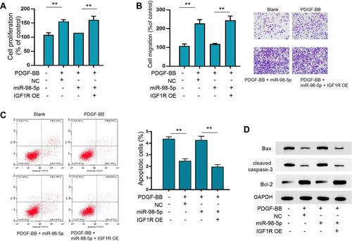
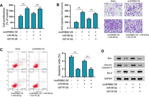
Figure 7 CircERBB2/miR-98-5p/IGF1R axis regulated the proliferation, migration and apoptosis of ASMCs. ASMCs were transfected with circERBB2 overexpressing vectors (circERBB2 OE), circERBB2 OE + miR-98-5p, or circERBB2 OE + miR-98-5p + IGF1R OE. Then, cell proliferation (A), migration (B), and apoptosis (C) of ASMCs were determined by CCK-8, Transwell, and flow cytometry assay, respectively. (D) The expression of Bax, cleaved caspase-3, and Bcl-2 was measured by Western blot analysis. **p < 0.01.