Figures & data
Figure 1 Flow chart of the participants inclusion and exclusion for this study.
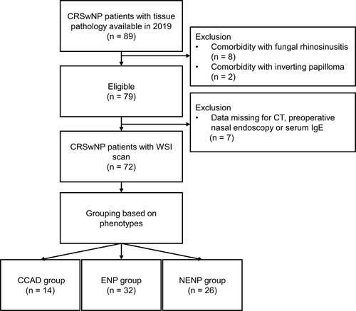
Table 1 Demographic Characteristics of Enrolled Patients
Figure 2 The endoscopic characteristics of CCAD, ENP, and NENP. (A), Middle turbinate polyposis in CCAD; (B and C) middle meatus polyps in ENP (B) and NENP (C).
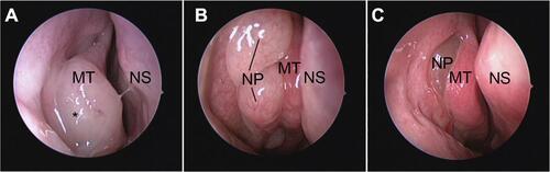
Figure 3 The radiographic features of CCAD, ENP and NENP. Note the central focus pattern of diseased sinus in central compartment atopic disease. General CT findings in CCAD (A), ENP (B), and NENP (C). Mucosal thickening involves the medial wall and floor of frontal sinus (D), ethmoid sinus (G), maxillary sinus (J), and sphenoid sinus (M) in CCAD. Mucosal thickening shows a diffused pattern and involves the frontal sinus, ethmoid sinus, maxillary sinus, and sphenoid sinus in ENP (E, H, K, N) and NENP (F, I, L, O).
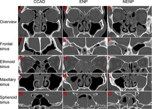
Figure 4 Comparison of aeroallergen prevalence of patients with CCAD, ENP, and NENP; *p < 0.05; **p < 0.01; ***p < 0.001.
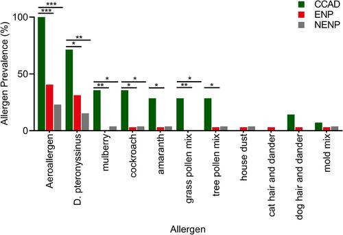
Figure 5 Cellular endotypes of CCAD, ENP, and NENP based on the WSI. (A-E) Representative photomicrographs of hematoxylin-eosin staining and heatmaps of eosinophil, neutrophil, lymphocyte and plasma cell in CCAD. (F-J) Representative photomicrographs of hematoxylin-eosin staining and heatmaps of eosinophil, neutrophil, lymphocyte and plasma cell in ENP. (K-O) Representative photomicrographs of hematoxylin-eosin staining and heatmaps of eosinophil, neutrophil, lymphocyte and plasma cell in NENP.
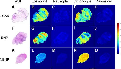
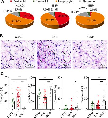
Figure 6 The cellular proportions in CCAD, ENP and NENP based on WSI. (A) The cellular endotypes in nasal tissues of different phenotypes of CRS. (B) Representative photomicrographs of hematoxylin-eosin staining (400 magnification) in CCAD, ENP, and NENP. Black arrows indicate representative eosinophils. (C) The proportions of eosinophil, neutrophil, lymphocyte and plasma cells in nasal tissues among groups. *P < 0.05, **P < 0.01, ***P < 0.001.