Figures & data
Table 1 Severity of Anaphylaxis
Figure 1 The month of the index sting. The number of enrolled patients who were stung by a vespid (orange, n=170) or bee (blue, n=25). The respective months are indicated on the x-axis. No bee or vespid stings that resulted in anaphylaxis were reported from December to February.
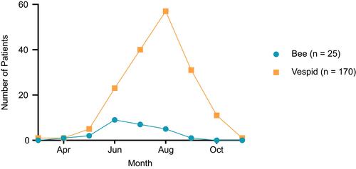
Figure 2 The severity of anaphylaxis and results of intracutaneous skin tests (ICTs). Values on the x-axis are severities of anaphylaxis graded I–IV according to Mueller.Citation15 ICTs were performed with increasing concentrations of vespid (left, orange, n=141) or bee (right, blue, n=13) venom until a positive reaction was seen. The colors within each of the bars indicate the venom concentration at which the ICT became positive in each group. Reless® (ALK-Abelló, Hamburg, Germany) and in rare cases Venomil® (Bencard, Munich, Germany) were used as allergen preparations.
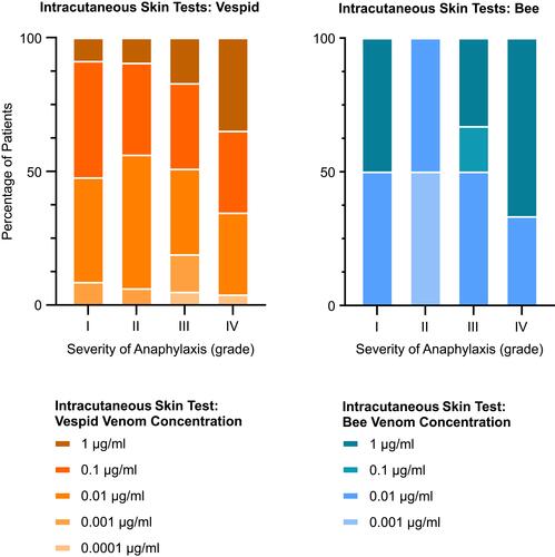
Figure 3 The severity of anaphylaxis, serum tryptase, and Sx1 levels. Values on the x-axis are severities of anaphylaxis Grades I–IV according to Mueller.Citation15 The horizontal lines indicate the arithmetic means. (A) Serum tryptase levels (µg/l) in individual patients with vespid (left, orange, n=117) or bee (right, blue, n=15) venom allergies. (B) Sx1 IgE (kU/l) levels in patients with vespid (left, orange, n=165) or bee (right, blue, n=25) venom allergies. The allergen mixture Sx1 was measured using the ImmunoCAP® system (Thermo Fisher, Waltham, MA, USA). Values below the diagnostic cut off according to the manufacturer’s instructions (0.35 kU/l for Sx1) were marked in red color. *Significant at α = 0.05.
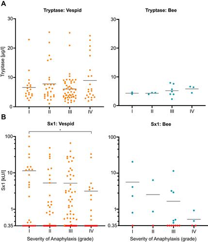
Figure 4 The severity of anaphylaxis and serum IgE levels. Values on the x-axis are severities of anaphylaxis Grades I–IV according to Mueller.Citation15 The horizontal lines indicate the arithmetic means. (A) Serum levels of venom-specific IgE (kU/l) in individual patients with vespid (left, orange, n=166) or bee (right, blue, n=25) venom allergies. (B) Serum levels of total IgE (kU/l) in individual patients with vespid (left, orange, n=168) or bee (right, blue, n=25) venom allergies. Values below the diagnostic cut off according to the manufacturer’s instructions (0.35 kU/l for both specific and total IgE) were marked in red color. *Significant at α = 0.05.
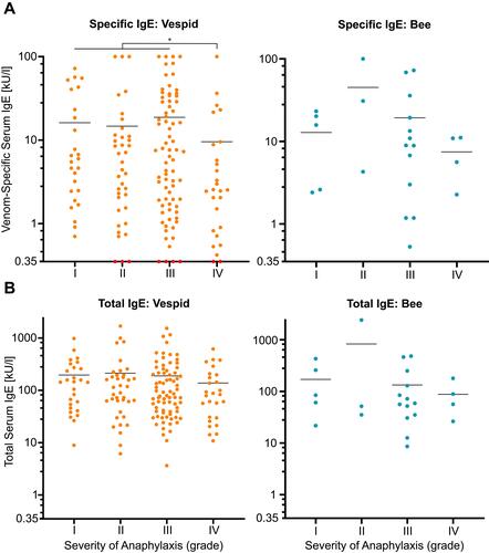
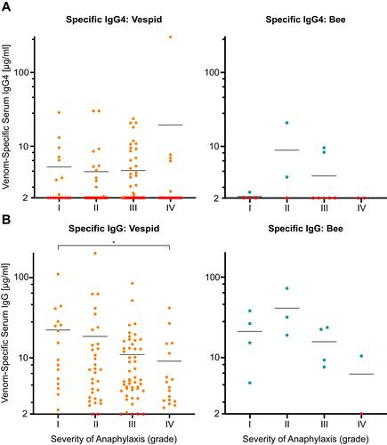
Figure 5 The severity of anaphylaxis and venom-specific serum IgG levels. Values on the x-axis are severities of anaphylaxis Grades I–IV according to Mueller.Citation15 The horizontal lines indicate the arithmetic means. (A) Serum levels of venom-specific IgG4 (µg/mL) in individual patients with vespid (left, orange, n=125) or bee (right, blue, n=16) venom allergies. (B) Serum levels of antigen-specific IgG (µg/mL) in individual patients with vespid (left, orange, n=116) or bee (right, blue, n=13) venom allergies. Values below the diagnostic cut off according to the manufacturer’s instructions (2 μg/mL for both IgG and IgG4) were marked in red color. *Significant at α = 0.05.