Figures & data
Figure 1 Decreased thickness of airway wall layers after BT as measured by endobronchial ultrasonography (EBUS).
Notes: (a) Representative EBUS image of the bronchus RB10 and the schematic outline of bronchial wall layers with major structural components highlighted. (b) The thickness of L1, L2 and L3–5 layers in asthma patients before BT and 12 months after the procedure. Data shown as medians and quartiles (n = 11; 2-sided exact Wilcoxon matched-pairs signed rank test).
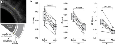
Table 1 Clinical Characteristics of Patients Before and After BT
Table 2 Correlation Between the Decrease in Thickness of Bronchial Layers and Other Parameters
Table 3 Histological Analysis of Bronchial Biopsies Before and After BT
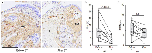
Figure 2 Bronchial wall histology changes related to BT.
Notes: (a) Representative histological images of bronchial biopsy samples showing reduction in airway smooth muscle (ASM) after BT at 12 months follow-up. α-SMA staining, hematoxylin-eosin counterstain; 100x magnification. Asterisk indicates fibrous connective tissue. (b) Reduction in the ASM (expressed as the percentage of cross-section surface area) before and after BT. (c) No change in the thickness of reticular basement membrane (RBM) after BT. Data shown as medians and quartiles (n = 11; 2-sided exact Wilcoxon matched-pairs signed rank test).