Figures & data
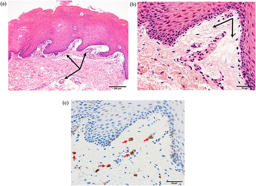
Figure 1 Buccal mucosal pathology after challenge testing. We biopsied our patient’s buccal mucosa immediately after intraoral bleeding developed due to her ingestion of potato snacks. Hematoxylin and eosin staining shows telangiectasia (black arrows) (a) and edema (black arrows) (b) of the subepithelial submucosa. Immunohistochemistry for c-kit reveals mast cells (red arrows) (c). The images in panels (b and c) are of the same section.