Figures & data
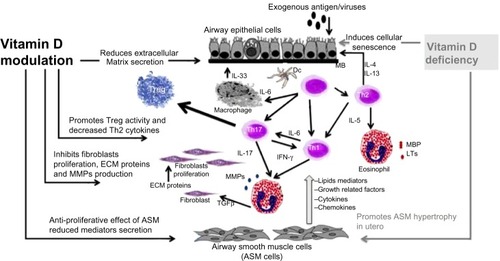
Figure 1 Potential pathways involving vitamin D in airway remodeling. Airway remodeling can be defined as changes in the composition, content, and organization of the cellular and molecular constituents of the airway wall. These structural changes include epithelial detachment, subepithelial fibrosis, increased ASM mass, goblet cell hyperplasia, mucous gland hyperplasia, and proliferation of blood vessels. Asthma exacerbation is triggered by allergens or viruses, inducing T-helper cell-driven inflammation and resulting in production of growth factors (TGF-β, TNF-α, and VEGF), profibrotic mediators (TGF-β, LTs, PG), MMPs, and ECM protein. Vitamin D insufficiency is associated with alteration in lung development, cellular senescence with inflammation and MMP release, and ASM hyperplasia. Potential beneficial effects of vitamin D are increasing Treg activity, improving innate immunity against viruses, decreasing release of induced mediators, and decreasing production of LTs, PG, MMP, TNF-α and TGF-β, resulting in decreased subepithelial fibrosis and ASM hypertrophy.