Figures & data
Figure 1 Histopathological features of hyaline-vascular type in a patient with multicentric Castleman’s disease. Left inguinal node biopsy shows atrophic germinal centers and expansion of the interfollicular zone with vascular proliferation (hematoxylin and eosin stain).
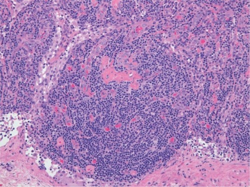
Figure 2 Proposed classification distinguishes multicentric Castleman’s disease on the basis of human herpesvirus 8 status.
Abbreviations: HHV8, human herpesvirus 8; HIV, human immunodeficiency virus; iMCD, idiopathic multicentric Castleman’s disease.
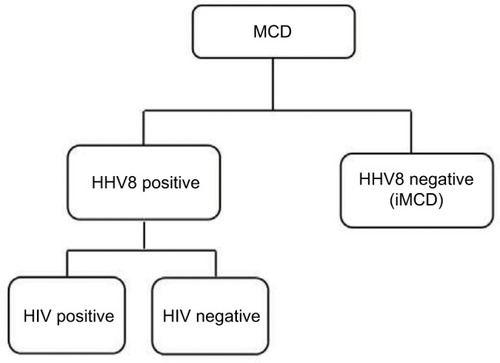
Table 1 Proposed diagnostic criteria for TAFRO-iMCD
Table 2 Definition of Castleman–Kojima disease (TAFRO syndrome)
Table 3 Consensus diagnostic criteria for iMCD
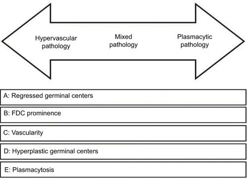
Figure 3 Histopathologic features of Castleman’s disease. Hypervascular subtype is characterized by the presence of regressed germinal centers and prominent FDC, whereas the plasmacytic subtype exhibits hyperplastic germinal centers and profuse plasmacytosis. The mixed subtype exhibits a combination of hypervascular and plasmocytic features. Vascularity is frequently observed in idiopathic multicentric Castleman’s disease but can be seen in either subtype as well. Republished with permission of American Society of Hematology, from International, evidencebased consensus diagnostic criteria for HHV-8 – negative/idiopathic multicentric Castleman disease, Fajgenbaum DC, Uldrick TS, Bagg A, et a, Blood, 129(12), 2017, permission conveyed through Copyright Clearance Center, Inc.Citation27