Figures & data
Figure 1 HbF response to HU is associated with hematological improvements in SCD patients.
Abbreviations: HbF, fetal hemoglobin; HU, hydroxyurea; SCD, sickle cell disease.
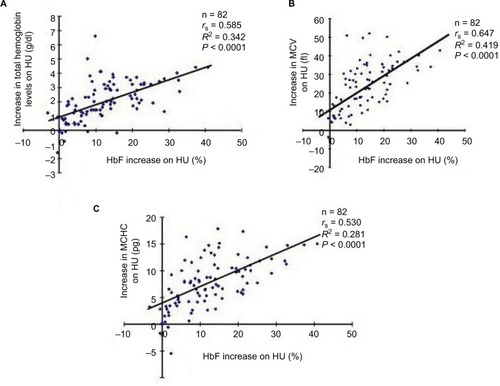
Figure 2 There were no correlations between HU-induced HbF increases and reductions in the number of reticulocytes (A) and platelets (B) among the SCD patients who received HU therapy.
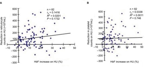
Figure 3 Correlations between the levels of HbF increases upon HU therapy and pre-HU therapy leukocyte counts (A), post-HU therapy leukocyte counts (B), or pre-HU therapy HbF levels (C). Eighty-two SCD patients who received HU therapy were analyzed.
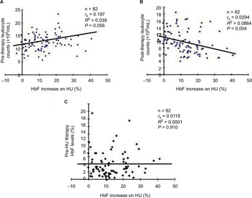
Figure 4 Correlations between neutrophil (A) or lymphocyte/monocyte counts (B) and HU-induced HbF levels.
Abbreviations: HU, hydroxyurea; HbF, fetal hemoglobin; SCD, sickle cell disease.
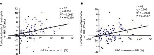
Figure 5 Presence of HbF silencing factors in serum of SCD patients.
Abbreviations: HbF, fetal hemoglobin; SCD, sickle cell disease; FBS, fetal bovine serum; GAPDH, glyceraldehyde 3-phosphate dehydrogenase; HU, hydroxyurea; GM-CSF, granulocyte-macrophage colony-stimulating factor; RT-PCR, real-time PCR.
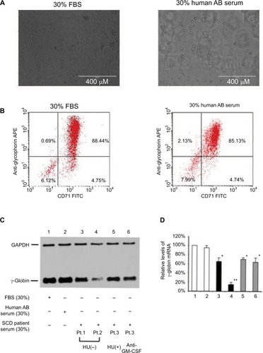
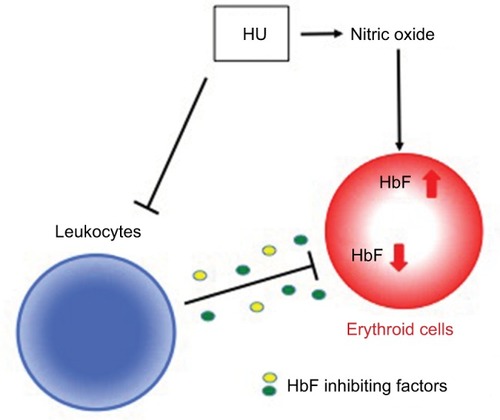
Figure 6 Possible model for the mechanisms that regulate HU-induced HbF expression.
Abbreviations: HU, hydroxyurea; HbF, fetal hemoglobin; sGC, soluble guanylate cyclase.