
Figures & data
Figure 1 Presumed mechanism of anti-platelet antibody and platelet destruction in ITP. A. Anti-GPIIb/IIIa antibody-coated platelets are trapped by the Fc receptors of activated macrophages, following which the platelets are destroyed. B. Activated macrophages present GPIIb/IIIa-derived antigen to T cells. C. These T cells interact with autoreactive B cells and present the GPIIb/III-derived antigen. D. Th1 cytokines such as IL-2 and IFNγ promote the activation of B cells. E. Anti-GPIIb/IIIa antibodies are produced by activated B cells. F. T-reg’s control is weakened.
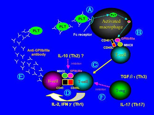
Table 1 Baseline Characteristics of the Patients with ITP and Healthy Controls
Table 2 Frequencies of Cytokine Gene Polymorphisms in ITP and Normal Controls
Table 3 Distribution of Cytokine Genotypes in Prednisolone (PSL) Responder and Nonresponder Patients with ITP
Table 4 Distribution of Cytokine Genotypes in Eltrombopag Responder and Nonresponder Patients with ITP
Table 5 Individual IL-10 Haplotype Frequencies and Genotypes in Prednisolone (PSL) or Eltrombopag (Elt) Responders and Nonresponder Patients with ITP