Figures & data
Table 1 Literature Review of Prior Cases with HLH Systolic Heart Failure
Table 2 Laboratory, Bone Marrow and ECHO Findings of Case 1 and 2
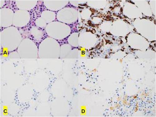
Figure 1 The bone marrow biopsy shows hypercellularity with increased megakaryocytes (A, H&E x40). The immunohistochemical studies show increased interstitial macrophages by CD68 (PGM1) (B, x40); and they are type 2 macrophages negative for pSTAT1 (C, x 0) but positive for CD163 (D, x40).
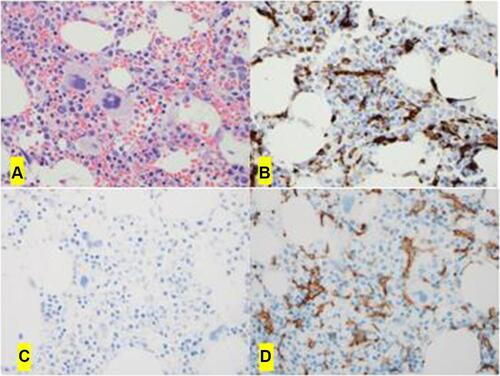
Figure 2 The bone marrow biopsy shows hypocellularity with left-shifted myelopoiesis (A, H&E x 0). Immunohistochemical studies shows increased interstitial macrophages by CD68 (PGM1) (B, x40); they are type 2 macrophages (M2) negative for pSTAT1 (C, x 0) and positive for CD163 (D, x40).