
Figures & data
Figure 1 Non-contrast CT scans.
Notes: (A) Normal liver and spleen. (B) Diffuse increased attenuation of the liver indicating excess iron deposition in a 47-year old patient with sickle cell disease undergoing multiple blood transfusions.
Abbreviation: CT, computed tomography.
Abbreviation: CT, computed tomography.
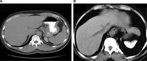
Figure 2 Normal axial T2* of liver of a 21-year old male patient with a known case of β-thalassemia on regular blood transfusions.
Notes: There is a rise in signal intensity as echo time increases. This patient had a normal liver iron concentration of 0.15 mg/g.
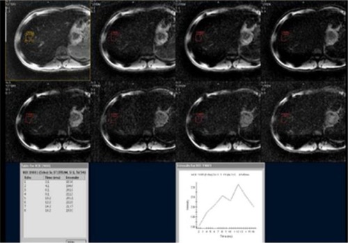
Figure 3 Axial T2* of liver and heart of a 24-year old female patient with a known case of β-thalassemia major on chronic blood transfusions.
Notes: (A) Moderate-to-high degree of hemosiderosis in this patient’s liver. T2* measurements of liver demonstrated sever hepatic iron loading of 2.1 ms. Sharp decrease in signal intensity as the echo time increases. Measured liver iron content was moderately high with 13.09 mg/g. (B) Heart – moderate degree of cardiac hemosiderosis demonstrating a gradual decrease in signal intensity as the echo time increases.
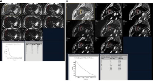
Table 1 Comparison of iron chelators