Figures & data
Figure 1 Schematic diagram of the experimental design.
Abbreviations: IP, intraperitoneal, IXD, Ixeris dentata; STZ, streptozotocin.
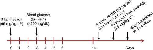
Figure 2 Effects of IXD extract on salivary parameters.
Notes: Saliva was collected for up to 30 min after pilocarpine injection and the salivary parameters were measured. (A) Total saliva collected in 30 min (mL). (B) Salivary flow rate (μL/min). (C) Bilateral submandibular gland weight (g). (D) Total salivary protein concentration (mg/mL). *significant difference vs vehicle-treated control rats ; #significant difference vs STZ-induced diabetic control rats (p<0.05). Values are represented as mean ± SEM (n=10 rats per group).
Abbreviations: IXD, Ixeris dentata; SEM, standard error mean; STZ, streptozotocin.
Abbreviations: IXD, Ixeris dentata; SEM, standard error mean; STZ, streptozotocin.
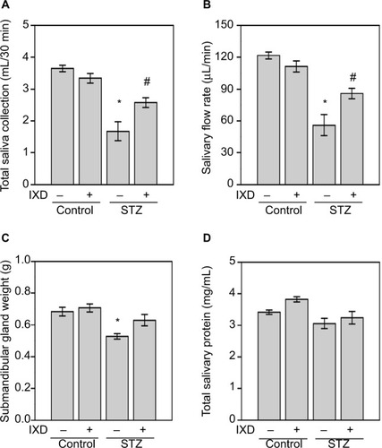
Figure 3 Effects of IXD extract on the morphology of submandibular glands and in salivary total protein expression.
Notes: (A) Hematoxylin and eosin staining was performed on paraffin-embedded submandibular gland tissues from normal and diabetic rats, either treated with water or IXD extract. Magnification=20×, scale bar=100 μm. (B) CBB staining showing total protein expression in the saliva of control and diabetic rats, either treated with water or IXD extract. The same volume of saliva was loaded in each well of a 10% SDS-PA gel, and the gel was stained with CBB. The black arrow indicates the molecular size of amylase (55 kDa) present in the saliva.
Abbreviations: CBB, Coomassie Brilliant Blue; IXD, Ixeris dentata; SDS-PAGE, sodium dodecyl sulfate polyacrylamide gel electrophoresis.
Abbreviations: CBB, Coomassie Brilliant Blue; IXD, Ixeris dentata; SDS-PAGE, sodium dodecyl sulfate polyacrylamide gel electrophoresis.
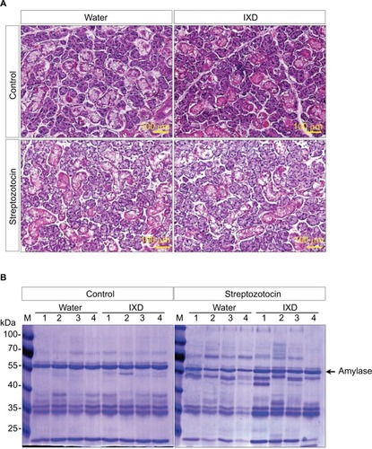
Figure 4 α-amylase protein expression was increased in IXD-treated diabetic rats
Notes: (A) Representative Western blot showing α-amylase expression in submandibular gland tissue homogenates. (B) Western blot showing α-amylase expression in saliva. (C) Immunohistochemical characterization showing the expression of α-amylase in submandibular gland tissue. The red/brown color indicates positive expression of α-amylase. Magnification=40×, scale bar=100 μm.
Abbreviations: IXD, Ixeris dentata; CBB, Coomassie Brilliant Blue.
Abbreviations: IXD, Ixeris dentata; CBB, Coomassie Brilliant Blue.
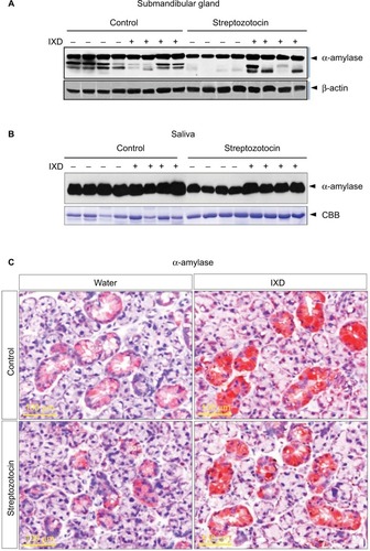
Figure 5 IXD extract increased AQP5 protein expression in submandibular gland tissue
Notes: (A) Representative Western blot showing AQP5 protein expression in SMG tissue homogenates. (B) Immunohistochemical analysis was performed on SMG tissue using anti-AQP5 antibody. The red/brown color indicates positive expression of AQP5. Magnification=40×, scale bar=100 μm.
Abbreviations: AQP5, aquaporin-5; IXD, Ixeris dentata; SMG, submandibular gland.
Abbreviations: AQP5, aquaporin-5; IXD, Ixeris dentata; SMG, submandibular gland.
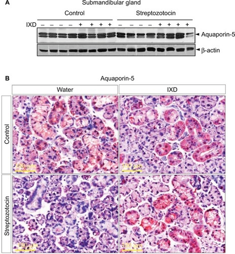
Figure 6 Confocal microscope images of immunofluorescence-detected α-amylase and AQP5 expression
Notes: Vehicle-or STZ-injected rats were treated with either water or IXD extract to observe the expression and localization of amylase and AQP5 in the submandibular gland. Double immunofluorescence staining was done using anti-amylase and anti-AQP5 antibodies. Green fluorescence indicates α-amylase expression, red fluorescence indicates AQP5 expression, and yellow fluorescence indicates co-localization of both proteins. Treatment with IXD extract produced a high intensity of amylase and AQP5 fluorescence compared with the controls. Magnification=40×, scale bar=1 μm.
Abbreviations: AQP5, aquaporin-5; STZ, streptozotocin; IXD, Ixeris dentata.
Abbreviations: AQP5, aquaporin-5; STZ, streptozotocin; IXD, Ixeris dentata.
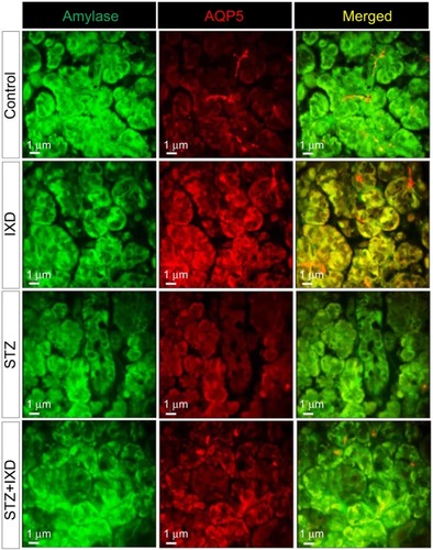
Figure 7 IXD extract treatment increased NHE1 expression in rat submandibular glands
Notes: Representative immunohistochemical detection of NHE1 in submandibular gland tissue from normal and diabetic rats, which were treated with either water or IXD extract. The yellow arrows indicate NHE1 expression in duct cells and the black arrows indicate NHE1 expression in acinar cells. Magnification=20×, scale bar=100 μm.
Abbreviations: NHE1, sodium/hydrogen exchanger-1; IXD, Ixeris dentata.
Abbreviations: NHE1, sodium/hydrogen exchanger-1; IXD, Ixeris dentata.
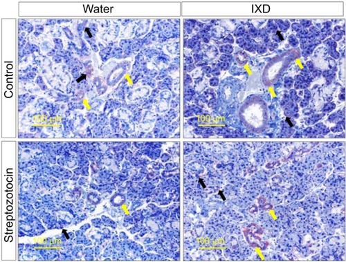
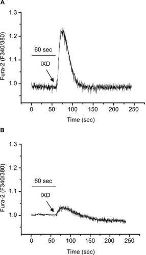
Figure 8 IXD extract increases intracellular calcium release in the HSG cells
Notes: (A) Fura-2-loaded HSG cells were promptly treated with IXD extract (0.02 mg/mL) and changes in F340/F380 were monitored. (B) The effect of 10 μM BAPTA-AM in IXD induced Ca2+ release. The figures represent the typical Ca2+ transients from more than three experiments.
Abbreviations: BAPTA-AM, 1, 2-Bis (2-aminophenoxy) ethane-N, N, N′, N′-tetraacetic acid tetrakis (acetoxymethyl ester); Ca2+, calcium; HSG, human salivary gland; IXD, Ixeris dentata.
Abbreviations: BAPTA-AM, 1, 2-Bis (2-aminophenoxy) ethane-N, N, N′, N′-tetraacetic acid tetrakis (acetoxymethyl ester); Ca2+, calcium; HSG, human salivary gland; IXD, Ixeris dentata.