Figures & data
Figure 1 a1) A strong positive reaction with tissue-type plasminogen activator (tPa) antibody was present in Chinese hamster ovary cells transfected with tPa plasmid (immunofluorescence 400×). a2) A negative reaction with tPa antibody in the control group (immunofluorescence 400×). b) The results of gel electrophoresis. c) Microbubbles under 400× microscope.
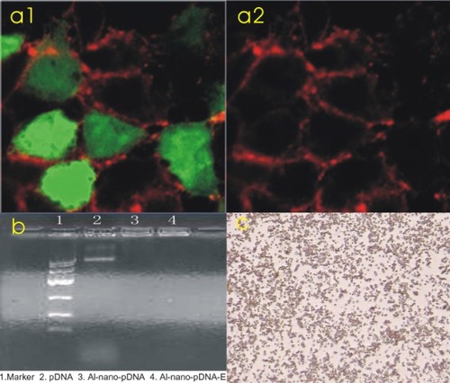
Figure 2 a1) A normal heart ultrasound image before injection of the microbubbles. a2) An increased heart ultrasound image after injection of microbubbles. a3) A heart ultrasound image after the treatment to return it to normal. b1) A normal liver ultrasound image before injection of the microbubbles. b2) An increased liver ultrasonic image after injection of the microbubbles. b3) A liver ultrasound image after the treatment to return it to normal.
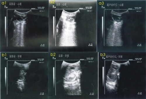
Figure 3 a1) Tissue-type plasminogen activator (tPa)-positive myocardium treated with ultrasound microbubbles (immunohistochemical stain 200×). a2) tPa-negative myocardium treated without ultrasound microbubbles (immunohistochemical stain 200×). b) tPa-positive skeleton muscles after transfection (immunohistochemical stain 200×) c) tPa-positive liver cells after transfection (immunohistochemical stain 400×). d) tPa-positive chondrocytes in the cartilage germinal layer after transfection (immunohistochemical stain 400×). e) tPa-positive myeloid cells and interstitial cells in medulla ossium after transfection (immunohistochemical stain 400×).
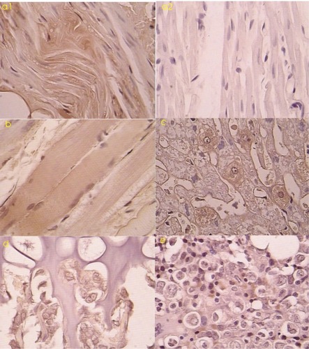
Table 1 Blood content of D-dimer and tissue-type plasminogen activator (tPA) before and after the targeting transgene (µg/L, x ± s)