
Figures & data
Table 1 Ethanol-Induced Gastric Ulcer Model; Treatment in Each Group
Table 2 Effects of DLBS2411 and Sucralfate on Ethanol-Induced Gastric Ulcers in Rats
Figure 1 Pathology of rats’ stomachs after being induced with ethanol. (A) Normal control group; (B) Negative control group; (C) Positive control group (100 mg/kg BW sucralfate); (D) 25 mg/kg BW DLBS2411; (E) 50 mg/kg BW DLBS2411. The arrows (►) show representative findings of gastric ulcers.
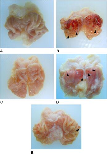
Figure 2 Classification of ulcer area.
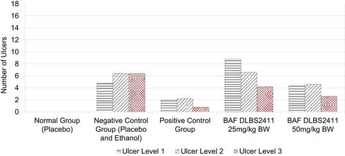
Figure 3 Histopathological examination of rats’ stomachs. Samples were from the stomach of a normal rat (A), a rat with the gastric ulcer treated with saline (B), a rat with the gastric ulcer treated with 100 mg/kg BW sucralfate (C), and rats with the gastric ulcer treated with 25 mg/kg BW DLBS2411 (D) and 50 mg/kg BW DLBS2411 (E). The slices were stained with hematoxylin and eosin and then examined under an optical microscope (20×). Mucosal desquamation (white arrows); necrosis of gastric mucosa (yellow arrows); undamaged gastric mucosal architecture (arrowheads).
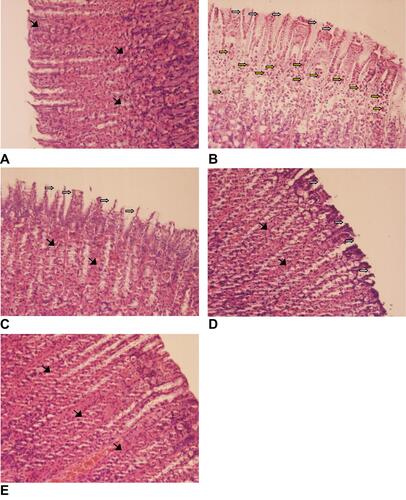
Table 3 Hematological Examination After Treatments