
Figures & data
Figure 1 Flowchart of the study cohort.

Table 1 Clinical and Pathologic Characteristics of Study Patients
Table 2 Imaging Characteristics of Hepatic Tumors in Cirrhotic Liver at Gadolinium Diethylenetriamine Pentaacetic Acid–Enhanced MRI
Table 3 Results of LI-RADS Categorization of 432 Hepatic Tumors
Table 4 Univariable and Multivariable Analyses of Clinical Factors Affecting Overall Survival and Recurrence-Free Survival
Figure 2 Recurrence-free survival and overall survival of 432 patients after surgical resection of primary liver cancer according to the Liver Imaging Reporting and Data System (LI-RADS) category and pathologic diagnosis. (a and b) Kaplan–Meier curves for overall survival (a) and recurrence-free survival (b) for patients with HCC, iCCA and HCC-CC. (c and d) Kaplan–Meier curves for overall survival (c) and recurrence-free survival (d) in the LR-M and LR-4/5 categories.
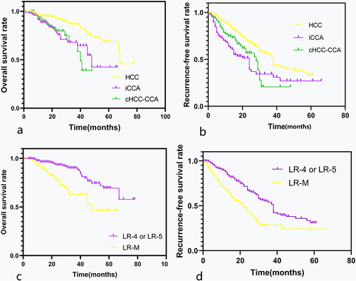
Figure 3 Recurrence-free survival and overall survival in patients with single primary liver cancer categorized as LR-M with tumor size >30 mm. Kaplan–Meier curves for overall survival (a) and recurrence-free survival (b) in LR-M with tumor size >30 mm according to pathologic diagnosis.
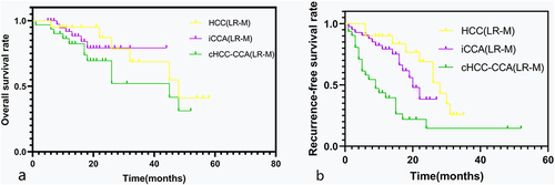
Figure 4 Recurrence-free survival and overall survival in patients with combined hepatocellular carcinoma-cholangiocarcinoma (cHCC-CCA) according to the Liver Imaging Reporting and Data System (LI-RADS) category in comparison with patients categorized as LR-4/5 with hepatocellular carcinoma (HCC) and patients categorized as LR-M with intrahepatic cholangiocarcinoma (iCCA). Kaplan–Meier curves show (a) overall survival and (b) recurrence-free survival after surgical resection of primary liver cancer.
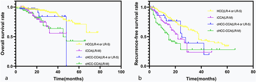
Figure 5 A 64-year-old male patient with hepatitis B–related liver cirrhosis and cHCC-CCA is categorized as Liver Imaging Reporting and Data System category LR-M. A 3.5 cm tumor in the right lobe of the liver shows heterogeneous hyperintensity on T2-weighted imaging (a) and diffusion weighted imaging (b). (c) The arterial phase shows peripheral enhancement on contrast-enhanced T1-weighted imaging with the contrast agent Gd-DTPA, and the nodule exhibits peripheral washout (red arrow) and an enhancing capsule (white arrow) in the delayed phase (d). Small recurrent lesions were found 5 months after surgery in this patient.
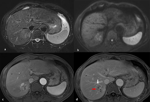
Figure 6 A 54-year-old male patient with hepatitis B–related liver cirrhosis and HCC is categorized as LR-M category. A 40-mm nodule shows heterogeneous hyperintensity on (a) diffusion weighted imaging. (b) T1-weighted imaging shows heterogeneous hypointensity in hepatic segment VIII. It presents rim arterial phase hyperenhancement (arrow) on contrast enhanced T1-weighted imaging (c) with contrast agent Gd-DTPA, resulting in delayed central enhancement (arrow) in (d) the delayed phase.
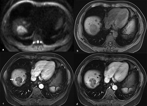
Table 5 Univariable and Multivariable Analyses of Imaging Features Affecting Overall Survival and Recurrence-Free Survival