Figures & data
Table 1 Characteristics of Patients with Unresectable Hepatocellular Carcinoma
Figure 1 Flow chart of the data selection process.
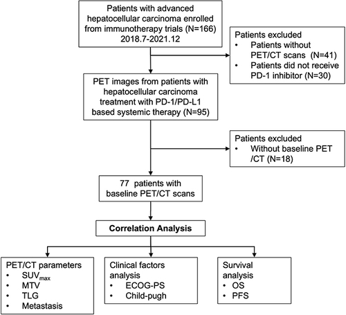
Figure 2 Kaplan–Meier curves for overall survival in relation to (A) SUVmax, (B) MTV, and (C) TLG (p values for Log rank test).
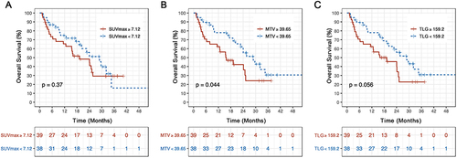
Table 2 Univariate Analysis of Prognostic Factors Associated with Overall Survival in Patients with Unresectable Hepatocellular Carcinoma
Table 3 Multivariate Analysis of Prognostic Factors Associated with Overall Survival in Patients with Unresectable Hepatocellular Carcinoma
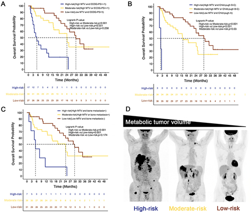
Figure 3 Kaplan–Meier curves for overall survival in three risk groups stratified according to MTV combined with the ECOG-PS (A), Child-Pugh grade (B), and bone metastasis (C). Illustration of high, moderate, and low risk using maximal intensity projection on 18F-FDG PET images of three patients (D).