
Figures & data
Figure 1 Flow chart of the study participants.
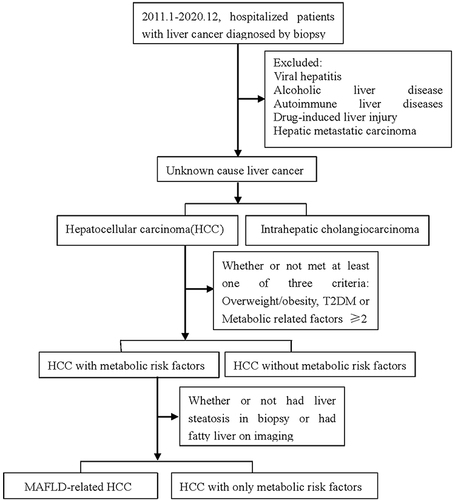
Figure 2 The degrees of surrounding nontumoral liver tissue steatosis in MAFLD-related HCC with liver cirrhosis background (A) and non-cirrhosis background (B), which are less than 30% (hematoxylin and eosin stain, original magnification ×100).
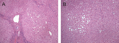
Table 1 Comparison Between Different Groups
Figure 3 HCC with metabolic risk factor architectural growth patterns: (A) The tumor cells grow in trabecular pattern. (B) The tumor cells grow in a macrotrabecular pattern with thick trabeculae more than 10 cell layers. (C) The tumor cells grow in a pseudoglandular pattern with glandular or acinar structure. (D) The tumor cells grow in a solid pattern without trabecular or pseudoglandular growth. (hematoxylin and eosin stain, original magnification ×100).
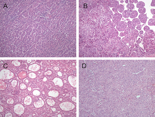
Figure 4 Major subtypes of HCC with metabolic risk factors. (A) Steatohepatitic HCC: The tumor shows macrovesicular steatosis, ballooning, Mallory–Denk bodies, and inflammation (hematoxylin and eosin stain, original magnification ×100). (B) Scirrhous HCC: The tumor nests and single tumor cells are present in a dense desmoplastic background (hematoxylin and eosin stain, original magnification ×200). (C) Clear-cell HCC: The tumor cells have glycogen-rich clear cytoplasm (hematoxylin and eosin stain, original magnification ×200).
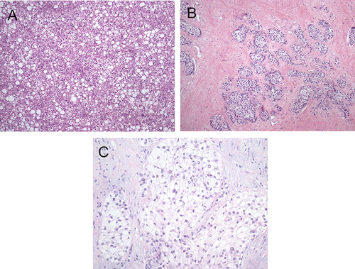
Figure 5 Histological features of steatohepatitic HCC. (A) Tumor cells with a group of ballooning hepatocytes with enlarged, rounded, rarefied cytoplasm, with some hepatocytes containing Mallory–Denk bodies (marked by arrow) (hematoxylin and eosin stain, original magnification ×400). (B) Glycogenated nuclei can be seen in some tumor cells (marked by arrow) (hematoxylin and eosin stain, original magnification ×200).
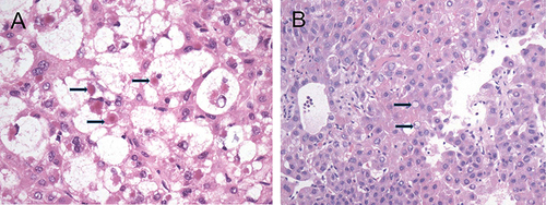
Table 2 Pathological Characteristics and Laboratory Indicators Between Different Pathological Types of HCC with Metabolic Risk Factors
Table 3 Correlation Analysis Between Pathological Characteristics and Laboratory Indicators of HCC with Metabolic Risk Factors
Table 4 Comparison of Liver Cirrhosis and Non-Cirrhosis Patients with HCC with Metabolic Risk Factors