Figures & data

Table 1 Clinical and Pathological Characteristics of MVI-Positive and MVI-Negative Cohorts
Figure 1 Flowchart showing the patient inclusion and exclusion criteria.
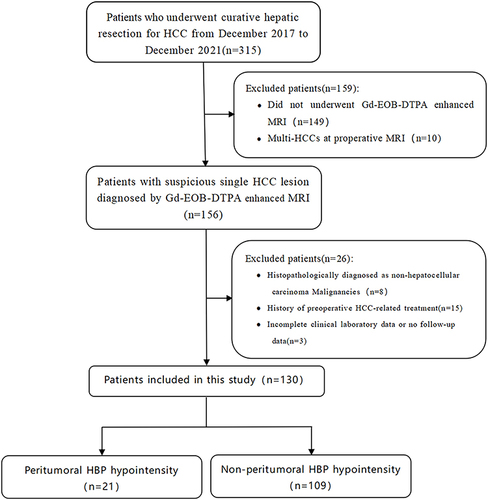
Table 2 The Interobserver Agreement of MRI Features
Table 3 MRI Features of MVI-Positive and MVI-Negative HCCs
Figure 2 Representative MRI features associated with histopathological findings. HBP images (a) for a 29-year-old male patient showing peritumoral HBP hypointensity (arrow) and satellite nodule (arrowhead). Hepatocellular carcinoma with MVI-positive was confirmed by histopathology (b). HBP images (c) for a 41-year-old male patient showing a radiological capsule (arrow) without peritumoral HBP hypointensity. Hepatocellular carcinoma with MVI-positive was confirmed by histopathology (d).
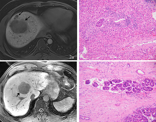
Table 4 MRI Features of MVI-Negative and MVI-Positive HCCs in Subgroup Without Peritumoral HBP Hypointensity
Figure 3 Kaplan–Meier curves showing recurrence-free survival in patients with and without peritumoral hepatobiliary phase hypointensity.
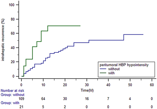
Table 5 Univariate and Multivariate Cox Regression of MRI Features in Predicting Intrahepatic Recurrence in Subgroup Without Peritumoral HBP Hypointensity
Figure 4 Kaplan–Meier curves showing recurrence-free survival in patients without peritumoral HBP hypointensity (PHBPH) and satellite nodule (SN), with SN but without PHBPH and with PHBPH.
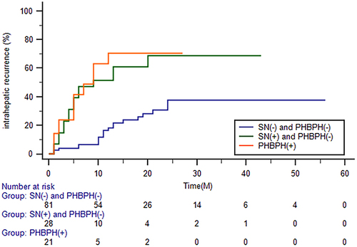
Figure 5 Representative MRI features associated with recurrence-free survival. HBP images (a) for a 60-year-old female patient showing an irregularly hypointense tumor (arrow) but without peritumoral HBP hypointensity or a satellite nodule. Tumor recurrence has not been found after 28 months of follow-up. HBP images (b) for a 50-year-old male patient showing multiple satellite nodules (arrow) around the tumor without peritumoral HBP hypointensity. Tumor recurrence was detected 4 months after curative resection.
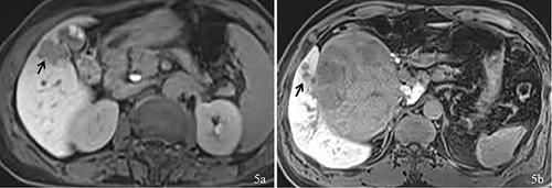
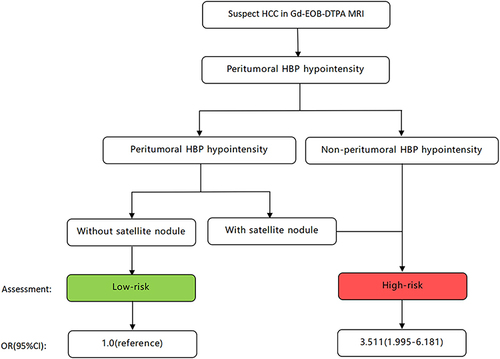
Figure 6 Two stepwise flowchart incorporating peritumoral hepatobiliary phase hypointensity and satellite nodule for stratification of the risk of intrahepatic recurrence. Recurrence-free survival was significantly shorter in patients with high-risk hepatocellular carcinoma than in those with low-risk hepatocellular carcinoma (hazard ratio 3.511, 95% confidence interval 1.995–6.181, P<0.001).