Figures & data
Figure 1 Imaging studies before and after ICI therapy. (A and B) A 4.83×3.73 cm hypervascular mass at liver S5. The mass had invaded her gallbladder and encased the right hepatic artery. (A) Arterial phase, (B) delayed phase; (C and D) after nivolumab, the tumor decreased from 4.83 cm to 3.21 cm (>30% reduction). The major vessels including right hepatic artery were spared from tumor invasion. (C) Arterial phase, (D) delayed phase.
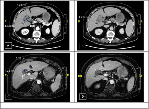
Figure 2 Imaging studies before and after ICI therapy. (A) A 10.32×7.15 cm hypervascular tumor was found at S6 of liver. (B and C) Recurrent HCCs were found at S6 (1.29 cm) (B) and S1 (2.82 cm) (C) of liver. (D) The tumor at S1 demonstrated partial response after 8 cycles (from 2.82 cm to 1.94 cm). However, it enlarged at cycle 13 (from 1.94 cm to 2.43 cm).
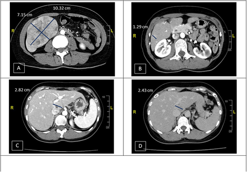
Figure 3 Imaging studies before and after ICI therapy. (A and B) A huge conglomerate tumor (18x14x10 cm) at the right inferior lobe of liver with obliteration of right portal vein. (C and D) The tumor reduced to 9.44×5.66 cm in size after 17 cycles of Atezo/Bev, and the right portal vein tumor thrombus (PVTT) also regressed.

Figure 4 Neoadjuvant therapy with ICI or Atezo/Bev combination.
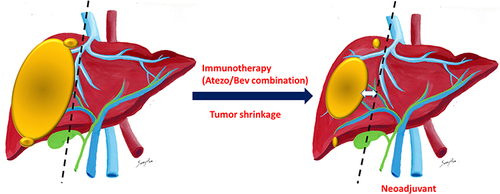
Figure 5 Negative selection by ICI or Atezo/Bev combination.
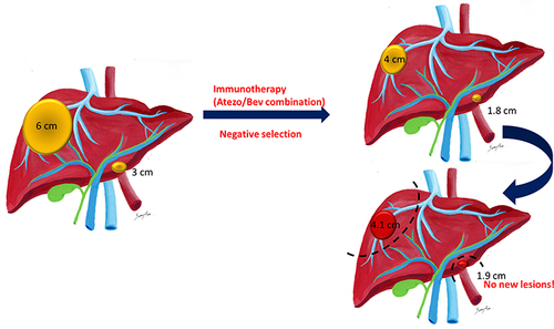
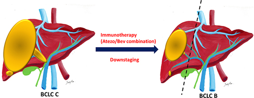
Figure 6 Downstaging by ICI or Atezo/Bev combination.