
Figures & data
Figure 1 A 22-year-old man with fibrolamellar HCC.
Notes: (A) Axial contrast-enhanced (gadoxetate disodium, Eovist, Bayer HealthCare), T1-weighted MR image obtained 20 minutes after contrast administration shows large multifocal fibrolamellar hepatocellular carcinoma with satellite lesions (arrows). (B) Coronal maximum-intensity projection image of 18F-FDG PET/CT shows that tumor has heterogeneous FDG avidity, with some lesions more FDG avid (arrow) than others (arrowhead). Reprinted with permission from the American Journal of Roentgenology.Citation50
Abbreviations: HCC, hepatocellular carcinoma; MR, magnetic resonance; PET, positron emission tomography; CT, computed tomography; FDG, fludeoxyglucose.
Abbreviations: HCC, hepatocellular carcinoma; MR, magnetic resonance; PET, positron emission tomography; CT, computed tomography; FDG, fludeoxyglucose.
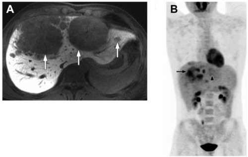
Figure 2 Typical histological features of FLHCC.
Notes: Hematoxylin and eosin staining of tumor tissues (A and B) from a patient with FLHCC. Large tumor cells are filled with eosinophilic granular cytoplasm and contain a large vesicular nucleus with a macronucleolus. They are arranged in trabeculae separated by abundant fibrous bands. (A) Low magnification; (B) high magnification. By Masson’s trichrome stain (C), dense fibrous bands (greenish blue) between nests of tumor cells are clearly visible. By immunohistochemical staining (D), tumor cells are strongly positive for Hepar, indicating the hepatocellular origin of tumor cells. Original magnifications are ×200 (A, C and D) and ×400 (B). Reprinted by permission from Macmillan Publishers Ltd: The American Journal of Gastroenterology. Liu S, Chan KW, Wang B, Qiao L. Fibrolamellar hepatocellular carcinoma. Am J Gastroenterol. 2009;104(10):2617–2624. Copyright © 2009.Citation56
Abbreviation: FLHCC, fibrolamellar hepatocellular carcinoma.
Abbreviation: FLHCC, fibrolamellar hepatocellular carcinoma.
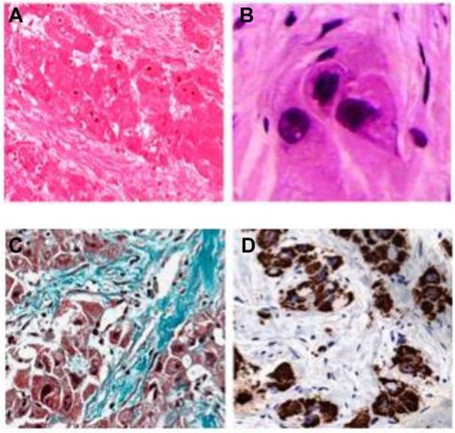
Figure 3 Overall survival of patients with FLC or HCC managed with a liver-directed procedure from the time of diagnosis in SEER from 1986 to 2008.
Notes: FLC (n=90; median survival: 75 months). HCC (n=7,135; median survival: 43 months). P=0.001. Reprinted from Journal of the American College of Surgeons, 218(2), Mayo SC, Mavros MN, Nathan H, et al, Treatment and prognosis of patients with fibrolamellar hepatocellular carcinoma: a national perspective, 196–205, Copyright © 2014, with permission from Elsevier.Citation70
Abbreviations: FLC, fibrolamellar carcinoma; HCC, hepatocellular carcinoma; SEER, Surveillance, Epidemiology, and End Results Program.
Abbreviations: FLC, fibrolamellar carcinoma; HCC, hepatocellular carcinoma; SEER, Surveillance, Epidemiology, and End Results Program.
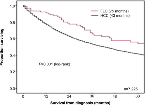
Table 1 Prognostic factors in fibrolamellar carcinoma