Figures & data
Figure 1 Mouse CAF1 gene knockout abolishes CAF1 protein expression.
Notes: Western immunoblot band density data showed that CAF1 gene knockout greatly reduced CAF1 levels and CAF1 band was nearly undetectable in CAF1-knockout mice. Western immunoblot band density data are expressed as mean ± SEM. n=3, #P<0.05 vs wild-type mice. An independent sample t-test was used.
Abbreviations: WT, wild-type mice; KO, CAF1-knockout mice; ns, normal saline; LPS, lipopolysaccharide; SEM, standard error of the mean.
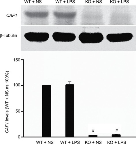
Figure 2 Effect of LPS on lung histology (40×).
Notes: Wild-type and CAF1-knockout mice challenged with ns displayed normal lung histology (A) and (B). On the contrary, lungs from wild-type mice stimulated with LPS exhibited features consistent with acute diffuse lung inflammation (C), which included alveolar and interstitial fluid accumulation, thickened alveolar wall, fibrin effusion, the infiltration of lymphocytes and neutrophils, and the destruction of pulmonary alveoli. These changes were more severe in LPS-stimulated CAF1-knockout mice (D).
Abbreviations: WT, wild-type mice; KO, CAF1-knockout mice; NS, normal saline; LPS, lipopolysaccharide.
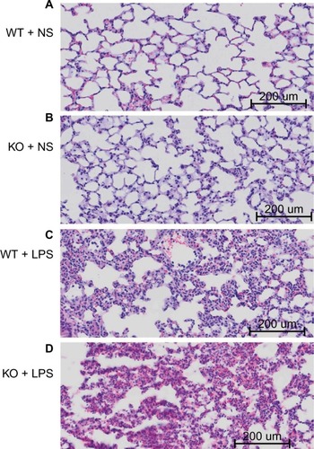
Figure 3 CAF1 knockout increases LPS-induced MPO activity.
Notes: MPO test data showed that MPO activity in CAF1-knockout mice treated with ns were comparable to those in wild-type mice. It indicated a significant increase in MPO activity both in wild-type and in CAF1-knockout mice following LPS stimulation, and MPO activity in CAF1-knockout mice was significantly higher than that in wild-type mice. The data are expressed as mean ± SEM. n=3, *P<0.05 vs. NS, #P<0.05 vs. wild-type mice. An independent sample t-test was used.
Abbreviations: WT, wild-type mice; KO, CAF1-knockout mice; ns, normal saline; LPS, lipopolysaccharide; MPO, myeloperoxidase.
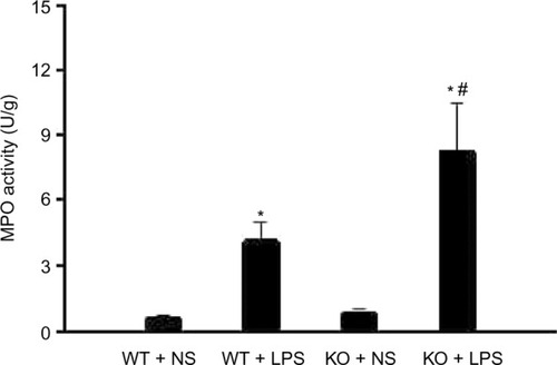
Figure 4 CAF1 knockout increases LPS-induced IL-8 and ICAM-1 protein levels.
Notes: ELISA data indicated IL-8 levels in CAF1-knockout mice challenged with ns were comparable to those in wild-type mice (A). It revealed a great increase in IL-8 both in wild-type and CAF1-knockout mice following LPS stimulation, and IL-8 levels in CAF1-knockout mice were greatly higher than those in wild-type mice (A). Western immunoblot band density data showed that ICAM-1 levels in wild-type mice challenged with ns were comparable to those in CAF1-knockout mice (B). LPS stimulation greatly increased ICAM-1 expression in both wild-type and CAF1−/− mice compared to ns stimulation. ICAM-1 levels in CAF1−/− mice stimulated with LPS were greatly more than those in LPS-stimulated wild-type mice (B). ELISA and Western immunoblot band density data are expressed as mean ± SEM. n=3, *P<0.05 vs NS, #P<0.05 vs wild-type mice. An independent sample t-test was used.
Abbreviations: WT, wild-type mice; KO, CAF1-knockout mice; ns, normal saline; LPS, lipopolysaccharide; ICAM-1, intercellular adhesion molecule-1; IL-8, interleukin-8; SEM, standard error of the mean; ELISA, enzyme-linked immunosorbent assay.
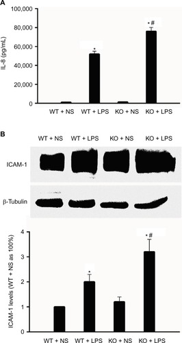
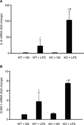
Figure 5 CAF1 knockout influences LPS-induced IL-8 and ICAM-1 mRNA expression. IL-8 and ICAM-1 mRNA levels in wild-type mice were comparable to those in CAF1−/− mice after NS stimulation and were greatly increased in both wild-type and CAF1−/− mice following LPS stimulation (A and B). IL-8 mRNA levels in CAF1−/− mice challenged with LPS were significantly higher than those in wild-type mice (A). ICAM-1 mRNA levels in CAF1−/− mice following LPS stimulation were significantly more than those in wild-type mice (B). The data are expressed as mean ± SEM. n=3, *P<0.05 vs NS, #P<0.05 vs wild-type mice. An independent sample t-test was used.(/P)(P)Abbreviations: WT, wild-type mice; KO, CAF1-knockout mice; ns, normal saline; LPS, lipopolysaccharide; ICAM-1, intercellular adhesion molecule-1; IL-8, interleukin-8; SEM, standard error of the mean.