Figures & data
Table 1 Medical conditions, involving SIJ
Figure 1 Early sacroiliitis.
Notes: Semicoronal MRI T2-weighted with fat saturation (A), T1-weighted (B) and semicoronal CT reconstruction (C) images of the SIJs of a 21 year old male with early sacroiliitis. BME is clearly seen on the right iliac side of the joint (arrowheads in A) as well as small erosions at the same location (arrowheads in B and C).
Abbreviations: BME, bone marrow edema; CT, computed tomography; MRI, magnetic resonance imaging; SIJ, sacroiliac joint.
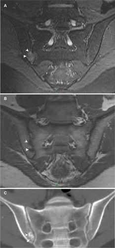
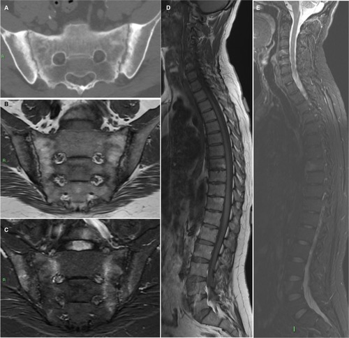
Figure 2 Forty two year old male with ankylosing spondylitis.
Notes: Axial CT image of the SIJs (A) demonstrating advanced disease with bilateral subchondral sclerosis, erosions, and pseudo-widening of the joints. Whole-spine MRI of the same patient with semicoronal T1-weighted (B), STIR (C) of the SIJs, and sagittal T1-weighted (D) and STIR (E) of the entire spine demonstrating bilateral SIJ’s BME, fat metaplasia, and erosions as well as corner inflammatory lesions and fatty lesions in the spine.
Abbreviations: BME, bone marrow edema; CT, computed tomography; MRI, magnetic resonance imaging; SIJ, sacroiliac joint; STIR, Short-TI inversion recovery.