Figures & data
Figure 1 Bacteriocin BacSp222 and its forms enhance NO production by P388.D1 and RAW 264.7 cells. (A and B) Determination of NO concentration measured by Griess reaction in culture media collected from the stimulated cells. (C and D) Analysis of the iNOS expression level in the stimulated cells using the qRT-PCR method. The bars represent the mean ± SEM (n=3). ***p<0.001 vs control.
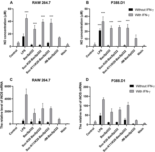
Figure 2 Activation of NF-κB in P388.D1 cells exposed to different forms of BacSp222. The cells were incubated for 30 minutes in the medium (control) and in the medium supplemented with LPS, BacSp222, suc-K20- BacSp222, or with -fM-BacSp222. The cells were then lysed, and the lysates were analyzed using the WB method. (A) Intracellular level of the p65 subunit in the cells. (B) Intracellular level of the phosphorylated p65 subunit detected using an antibody specific to phosphorylated p65 Ser536. The WB results are representative of three independent experiments.
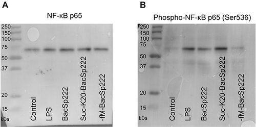
Figure 3 Production of selected cytokines by RAW 264.7 cells exposed to various forms of bacteriocin BacSp222. The cells were incubated in the control medium and in the medium supplemented with LPS, BacSp222, suc-K20-BacSp222, suc-K11/K20- BacSp222, -fM-BacSp222, or nisin for 24 h. After 24 h, the media were collected and subjected to flow cytometry analyses using a LEGEND/Plex Mouse Inflammation Panel kit to determine cytokine concentrations. The bars represent the mean ± SEM (n=8). ***p<0.001 vs control, **p<0.01 vs control, *p<0.05 vs control.
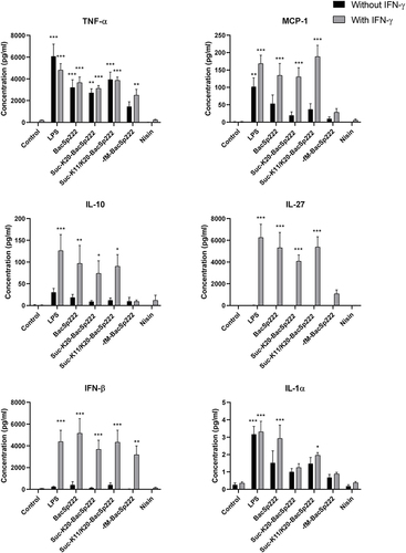
Figure 4 Analysis of TNFα secretion by murine monocyte-macrophage cells exposed to various forms of bacteriocin BacSp222. The P388.D1 (A) and RAW 264.7 (B) cells were incubated for 6 h in the control medium or in the medium with IFN-γ. Additionally, the media were supplemented with LPS, BacSp222, -fM-BacSp222, or nisin. The concentration of TNFα released by the cells to the culture media was determined using ELISA. (C) The P388.D1 cells were treated for 6 h with LPS to stimulate TNFα expression, and then the media were changed to remove proteins produced by the cells during the incubation. Next, the concentration of TNFα released from the cell membranes was analyzed in media collected from cells pretreated with metalloproteinase inhibitors (galardin, phenanthroline, 30 minutes) and then stimulated with PMA or BacSp222 and -fM-BacSp222 (45 minutes). The bars represent the mean ± SEM (n=3). ***p<0.001 vs control, **p<0.01 vs control, *p<0.05 vs control.
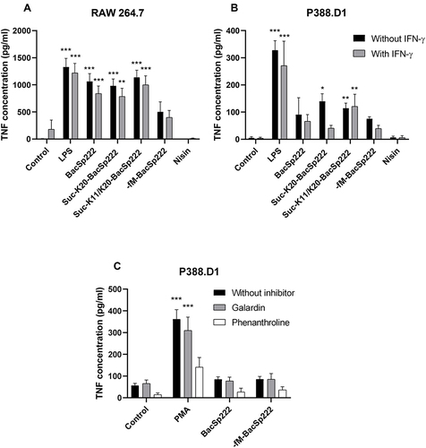
Figure 5 Bacteriocins BacSp222 stimulate IL-8 production by human PMNs. The cells were incubated overnight in control media or in media with LPS, BacSp222, suc-K20- BacSp222, or -fM-BacSp222. The concentration of IL-8 in the culture media was determined by the ELISA assay. The bars represent the mean ± SEM (n=3). ***p<0.001 vs control, **p<0.01 vs control.
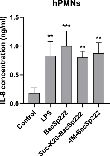
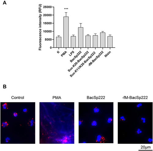
Figure 6 Analysis of ROS production by human PMNs exposed to the various forms of bacteriocins. Human PMNs were stained with ROS probes: DHE, DHR, or DCF-HDA and exposed to PMA or bacteriocins. Fluorescence intensity corresponding to the level of intracellular ROS was measured for 3 or 14 h, depending on the probe used (kinetic measurements). The graphs demonstrate the fluorescence intensity measured at the selected time points. The bars represent the mean ± SEM (n=3). ***p<0.001 vs control, **p<0.01 vs control, *p<0.05 vs control.
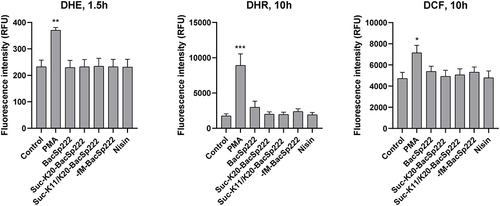
Figure 7 Bacteriocins do not stimulate NETs formation by human PMNs. (A) Human PMNs were incubated in the control medium or with PMA, LPS, various forms of bacteriocin BacSp222, or nisin for 4 h. Released cellular DNA was detected by fluorescence measurement after staining with SYTOX™ Green Nucleic Acid Stain. The bars represent the mean ± SEM (n=5). ***p<0.001. (B) Human PMNs were incubated in the control medium or with PMA, BacSp222, or -fM-BacSp222. The cellular DNA stained with DAPI (blue) and MPO stained using specific antibodies (red) were visualized using fluorescence microscopy.