Figures & data
Table 1 Primer Sequences
Figure 1 The chemical structure of engeletin and effects of engeletin on NP cells viability and proliferation. (A) The chemical structure of engeletin. (B) NP cells at a density of 2×10⁴ cells/well were treated with engeletin at different concentrations for 24h. CCK-8 tests were used to evaluate the cytotoxicity. (C–E) The influence of engeletin for proliferation was also measured by CCK-8 tests. NP cells at a density of 5×10³ cells/well were incubated with engeletin for 24h (C), 48h (D) or 72h (E). All data are presented as mean ± SD. *P <0.05, **P < 0.01, n = 3.
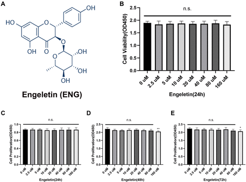
Figure 2 Engeletin ameliorates TNF-α-induced inflammation on NP cells. (A) The relative mRNA expression of iNOS, IL-1β, IL-6, COX2 and TNF-α were measured by RT-qPCR. (B and D) The expressions of iNOS, IL-1β and COX2 at protein level were determined by Western blotting. (C) The representative merge images of iNOS, IL-1β and COX2 were captured by immunofluorescence combined with DAPI staining for nuclei (scale bar: 100µm). (E) The results of immunofluorescence were analyzed through measuring the IOD values. All data are presented as mean ± SD. *P <0.05, **P < 0.01, ***P<0.001 and ****P<0.0001, n = 3.
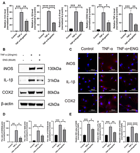
Figure 3 Engeletin prevented NP cells from the disturbance of metabolic homeostasis induced by TNF-α. (A) The relative mRNA expression of MMP3, MMP9, MMP13, SOX9, Collagen II and Aggrecan were analyzed by RT-qPCR. (B and D) The expression of MMP3, MMP9, MMP13 and SOX9 at protein level were detected by Western blotting. (C) The representative merge images of MMP3, MMP9 and Collagen II were obtained by immunofluorescence combined with DAPI staining for nuclei (scale bar: 100µm). (E) The results of immunofluorescence were analyzed through measuring the IOD values. (F) Alcian Blue staining for the high-density culture of NP cells for 1 week (scale bar: 2mm and 10µm). (G) The integrated optical density (IOD) value and IOD value/area were measured to evaluate the results of high-density culture through semi-quantitative analysis. All data are presented as mean ± SD. *P <0.05, **P < 0.01, ***P<0.001 and ****P<0.0001, n = 3.
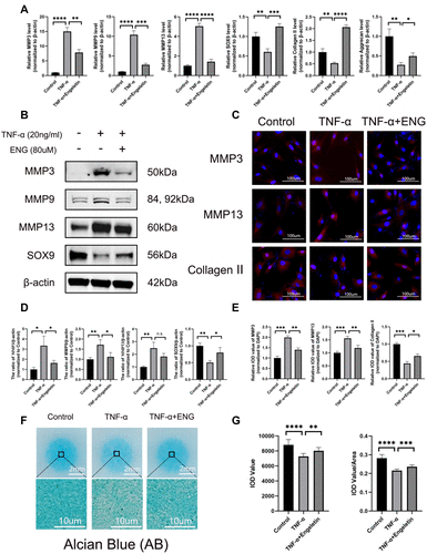
Figure 4 Engeletin alleviated TNF-α-induced apoptosis of NP cells. (A and D) Stained with Annexin V-FITC and PI, the rate of apoptotic NP cells in different groups were measured by flow cytometry. (B) The representative images of TUNEL assay with DAPI staining for nuclei (scale bar: 50µm). (E) TUNEL-positive cells were counted for the semi-quantitative analysis. (C and F) The expression of Bax, Bcl-2 and cleaved caspase-3 at protein level were determined by Western blotting. All data are presented as mean ± SD. **P < 0.01, ***P<0.001 and ****P<0.0001, n = 3.
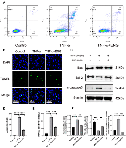
Figure 5 Engeletin down-regulated the TNF-α-induced NF-κB and MAPK signaling pathways activation in NP cells. (A and C) The expression of total IKKβ, IκBα, p65 and phosphorylated IKKα/β, IκBα, p65 at protein level were measured by Western blotting. (B and D) The expression of total JNK, ERK, p38 and phosphorylated JNK, ERK, p38 at protein level were measured by Western blotting. (E and F) The phosphorylation and nuclear translocation of p65 and p38 were detected by immunofluorescence (scale bar: 20µm). All data are presented as mean ± SD. *P <0.05, **P < 0.01, ***P<0.001 and ****P<0.0001, n = 3.
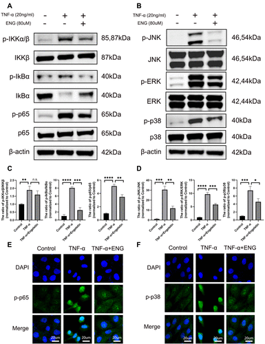
Figure 6 Engeletin ameliorated IDD in a rat model in vivo. (A) The representative X-ray and MRI images of a rat tail after 4 weeks of surgery. (B and C) The disc height index, Pfirrmann grades and histological scales were shown in different groups at 4 weeks. (E) The representative images of HE, SO-FG and AB staining of punctured IVD in different groups (scale bar: 500µm). (F and D) The representative images and semi-quantitative analysis of TUNEL staining for IVD tissues in different groups (scale bar: 25µm). (G) The expression of MMP13, MMP3, Collagen II, COX2, IL-1β and iNOS in IVD tissues at different groups were evaluated by immunohistochemistry staining (scale bar: 50µm). (H) The IOD value/area was calculated to assess the results of immunohistochemistry staining through semi-quantitative analysis. All data are presented as mean ± SD. *P <0.05, **P < 0.01, ***P<0.001 and ****P<0.0001, n = 3.
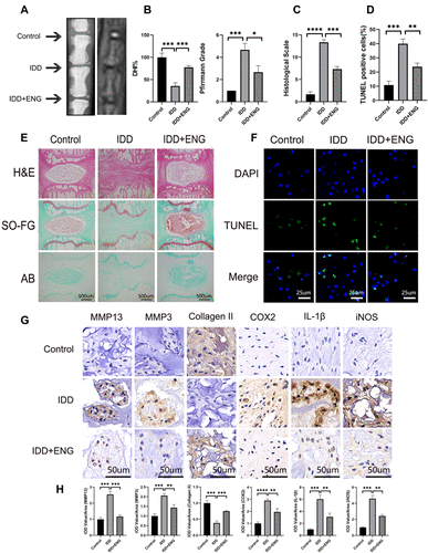
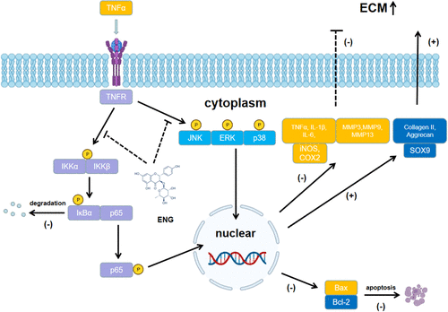
Figure 7 Potential molecular mechanism involved in the therapeutic effects of engeletin for IDD.