Figures & data
Figure 1 CEVs induced inflammatory response in NHEKs. (A) The morphology of CEVs (white arrow) was observed by TEM. Scale bar, 200 nm. (B) Cell viability of NHEKs 24 h after CEVs stimulation at different concentrations (0, 5, 10, 20, 50 μg/mL) was detected by CCK-8 assay. (C and D) mRNA levels of IL-6, IL-8 and TNF-α following CEVs stimulation at different concentrations (C) and different time points (D) were detected by RT-PCR. (E–G) Protein levels of IL-6, IL-8 and TNF-α in culture supernatants of NHEKs incubated with 10 μg/mL of CEVs for 12 h were detected by ELISA. The data are presented as the mean ± S.E.M. of three independent experiments. *P < 0.05; **P < 0.01; ****P < 0.0001.
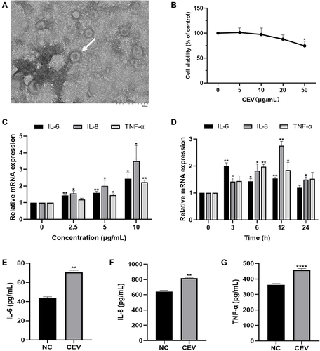
Figure 2 CBD suppressed the inflammatory reaction induced by CEVs in NHEKs. (A) Cell viability of NHEKs 24 and 48 h after CBD treatment at different concentrations (0, 1, 2, 5, 10, 20 μM) was detected by CCK-8 assay. (B–D) mRNA (B) and protein (C and D) levels of IL-6, IL-8 and TNF-α of CEVs-stimulated NHEKs in the presence of vehicle or CBD (0.5, 1 and 2 μM) were detected by RT-PCR (B), Western blotting (C, β-actin was used as the endogenous control) and ELISA (D) respectively. The data are presented as the mean ± S.E.M. of three independent experiments. *P < 0.05; **P < 0.01; ***P < 0.001; ****P < 0.0001.

Figure 3 The anti-inflammatory action of CBD was mediated by CB2 receptor and enhanced by the TRPV1 antagonist. (A–C) Protein levels of CB2 receptor and TRPV1 in the presence or absence of CEVs (10 μg/mL) and CBD (1 μM) were detected by Western blotting. β-actin was used as the endogenous control. (D) Cell viability of NHEKs co-stimulated with CBD (1 μM) and different concentrations (0, 1, 2, 5, 10, 20 μM) of AM630 or Capsazepine for 24 h was detected by CCK-8 assay. (E) TNF-α mRNA levels of NHEKs in the presence of AM630 or Capsazepine (0.01, 0.1, 1 and 5 μM) were detected by RT-PCR. The data are presented as the mean ± S.E.M. of three independent experiments. *P < 0.05; ****P < 0.0001 vs NC group; ##P < 0.01; ###P < 0.001 vs CEV group; &P < 0.05 vs CEV+CBD group.
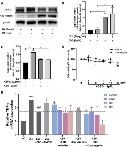
Figure 4 CBD inhibited activation of the MAPK and NF-κB signaling pathways. (A–C) The levels of MAPK- and NF-κB-related proteins of CEVs-stimulated NHEKs in the presence of vehicle or CBD (0.5, 1 and 2 μM) were detected by Western blotting. β-tubulin was used as the endogenous control. (D) Nuclear translocation of NF-κB p65 in CEVs-stimulated NHEKs in the presence of vehicle or CBD (1 μM) was detected by immunofluorescence staining. Scale bar, 100 μm. The data are presented as the mean ± S.E.M. of three independent experiments. *P < 0.05.
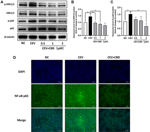
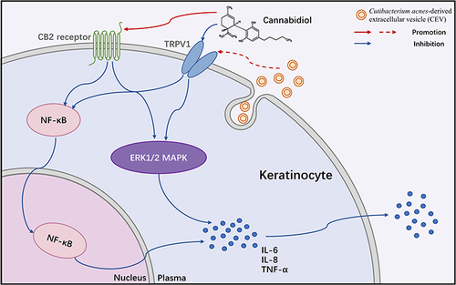
Figure 5 Mechanisms underlying the inhibitory effect of cannabidiol on acne inflammation induced by Cutibacterium acnes-derived extracellular vesicles in keratinocytes.