Figures & data
Table 1 Laboratory Finding on Admission
Figure 1 Left pelvic cyst-solid mass with ultrasound (size 16.3×9.7x7.6 cm). Criss-cross: left pelvic cyst-solid mass.
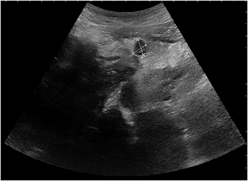
Figure 2 CT showed effusion of bilateral thoracic cavities (yellow arrow).
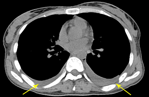
Figure 3 MRI showed abnormal signals of bilateral appendages, multiple small nodule shadows in greater omentum, lesser omentum and mesentery, abnormal signal shadows in pelvis, sacrum, thoracic vertebrae and lumbar vertebrae, effusion in abdominal, pelvic cavities. (A) T1WI, (B) T2WI, (C) STIR, (D) T1+ C. White arrow: iliac bone metastasis.
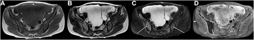
Table 2 Lymphocyte Subsets Results
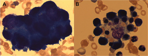
Figure 4 The bone marrow showed clustered abnormal cells (A) and haemophagocytes (B) (Wright-Giemsa staining, original magnification x 1000).