
Figures & data
Table 1 Correlation of Age and Gender with H. pylori Infection Developing to Intraepithelial Neoplasia
Figure 1 Pathological stages of the occurrence and development of H. pylori infection in gastric mucosa.
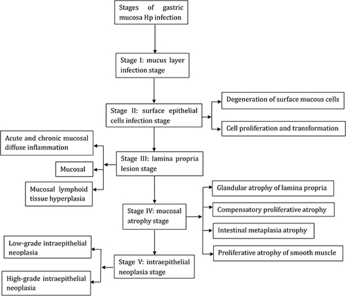
Table 2 The Histomorphological Characteristics, Immunophenotype, and Pathological Stages of Helicobacter pylori Infection in the Gastric Mucosa from Occurrence to Intraepithelial Neoplasia
Figure 2 Stage I, the mucus layer infection stage: (A) Helicobacter pylori (Hp) colonizes in the mucous layer covered by the epithelium on the surface of the gastric mucosa (hematoxylin and eosin staining, ×400); (B) the result for Hp is positive (EnVision method, × 400).
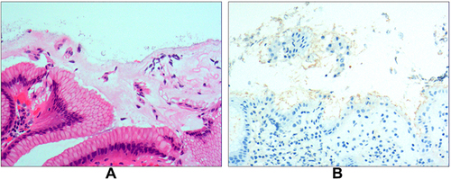
Figure 3 Stage II, surface epithelial cell infection. (A) Stage IIA, the degeneration of mucus surface epithelial cells: the amount of Helicobacter pylori (Hp) bacteria is significant, and the cytoplasm exhibits spiderweb-like vacuolar degeneration; the remaining cytoplasm surrounds the nucleus in a radial pattern, while a characteristic Hp-infection band can be observed under low magnification (hematoxylin and eosin staining [H&E], ×100). (B) Stage IIB, cell proliferation and transformation: I) A large amount of Hp adheres in the cytoplasm of the surface epithelial cells, resulting in massive proliferation and transformation of the cells in the depth of the gastric pit and the isthmus of the gastric gland, as well as in the proliferative zone in the upper part of the gland neck (H&E, ×200); II) the result for Hp is positive (EnVision method, ×400).
![Figure 3 Stage II, surface epithelial cell infection. (A) Stage IIA, the degeneration of mucus surface epithelial cells: the amount of Helicobacter pylori (Hp) bacteria is significant, and the cytoplasm exhibits spiderweb-like vacuolar degeneration; the remaining cytoplasm surrounds the nucleus in a radial pattern, while a characteristic Hp-infection band can be observed under low magnification (hematoxylin and eosin staining [H&E], ×100). (B) Stage IIB, cell proliferation and transformation: I) A large amount of Hp adheres in the cytoplasm of the surface epithelial cells, resulting in massive proliferation and transformation of the cells in the depth of the gastric pit and the isthmus of the gastric gland, as well as in the proliferative zone in the upper part of the gland neck (H&E, ×200); II) the result for Hp is positive (EnVision method, ×400).](/cms/asset/c9718f57-a5ee-485b-a579-4d62da953040/djir_a_12150028_f0003_c.jpg)
Figure 4 Stage III, the lamina propria lesion stage. (A) Stage IIIA involves acute and chronic diffuse mucosal inflammation throughout the lamina propria, the basophilic nature of the stroma in the intercellular space enhances, and a large number of infiltrated lymphocytes, plasma cells, neutrophils, and eosinophils exhibit different degrees of vacuolar degeneration (hematoxylin and eosin [H&E] staining, × 200). (B) Stage IIIB involves mucosal ulcers, with necrotic cells and tissue debris, as well as fibrous materials, small blood vessels, and inflammatory cells constituting the ulcers (H&E staining, ×100). (C) Stage IIIC involves mucosal lymphoid hyperplasia: I) diffuse small lymphocytic hyperplasia and various lymphoid follicles of different sizes; hyperplasia of the glands is rare (H&E staining, ×100); II) the result for Helicobacter pylori is positive (EnVision method, ×100).
![Figure 4 Stage III, the lamina propria lesion stage. (A) Stage IIIA involves acute and chronic diffuse mucosal inflammation throughout the lamina propria, the basophilic nature of the stroma in the intercellular space enhances, and a large number of infiltrated lymphocytes, plasma cells, neutrophils, and eosinophils exhibit different degrees of vacuolar degeneration (hematoxylin and eosin [H&E] staining, × 200). (B) Stage IIIB involves mucosal ulcers, with necrotic cells and tissue debris, as well as fibrous materials, small blood vessels, and inflammatory cells constituting the ulcers (H&E staining, ×100). (C) Stage IIIC involves mucosal lymphoid hyperplasia: I) diffuse small lymphocytic hyperplasia and various lymphoid follicles of different sizes; hyperplasia of the glands is rare (H&E staining, ×100); II) the result for Helicobacter pylori is positive (EnVision method, ×100).](/cms/asset/fe311051-48f9-4b53-830f-f992e25e9e5e/djir_a_12150028_f0004_c.jpg)
Figure 5 Stage IV, mucosal atrophy. (A) Stage IVA involves glandular atrophy of the lamina propria; the Helicobacter pylori-infected surface epithelial cells decrease, the surface mucus cells exhibit different degrees of proliferation and transformation, and the lamina propria glands are significantly smaller and decreased in number (hematoxylin and eosin [H&E] staining, ×200). (B) Stage IVB involves compensated proliferative atrophy; the lamina propria glands decrease or disappear and the surface epithelial cells exhibit compensatory proliferation (H&E staining, ×100). (C) Stage IVC involves intestinal metaplastic atrophy, where gland atrophy coexists with intestinal metaplasia; the glands of the intestinal metaplasia exhibit varying degrees of hyperplasia and expansion, as well as mild atypia (H&E staining, ×200). (D) Stage IVD involves smooth muscle proliferative atrophy: I) from the muscularis mucosa to the mucosal proliferation zone, proliferated smooth muscle forms a muscle fiber plate with characteristic fibrous tissue and smooth muscle together with the muscularis mucosa (H&E staining, ×200); II) the MUC6 expression is positive, exhibiting reduced glands of the fundus of the stomach (EnVision method, ×100).
![Figure 5 Stage IV, mucosal atrophy. (A) Stage IVA involves glandular atrophy of the lamina propria; the Helicobacter pylori-infected surface epithelial cells decrease, the surface mucus cells exhibit different degrees of proliferation and transformation, and the lamina propria glands are significantly smaller and decreased in number (hematoxylin and eosin [H&E] staining, ×200). (B) Stage IVB involves compensated proliferative atrophy; the lamina propria glands decrease or disappear and the surface epithelial cells exhibit compensatory proliferation (H&E staining, ×100). (C) Stage IVC involves intestinal metaplastic atrophy, where gland atrophy coexists with intestinal metaplasia; the glands of the intestinal metaplasia exhibit varying degrees of hyperplasia and expansion, as well as mild atypia (H&E staining, ×200). (D) Stage IVD involves smooth muscle proliferative atrophy: I) from the muscularis mucosa to the mucosal proliferation zone, proliferated smooth muscle forms a muscle fiber plate with characteristic fibrous tissue and smooth muscle together with the muscularis mucosa (H&E staining, ×200); II) the MUC6 expression is positive, exhibiting reduced glands of the fundus of the stomach (EnVision method, ×100).](/cms/asset/89172446-8c65-462c-948a-dc28290b253b/djir_a_12150028_f0005_c.jpg)
Figure 6 Stage V, intraepithelial neoplasia. (A) Stage VA involves low-grade intraepithelial neoplasia, where the glands exhibit irregular branching, glandular epithelial cells are present with mild-to-moderate atypia, the basophilic nature of the cytoplasm increases, the nuclei become longer, and the neoplasia is located at the base of the glandular epithelium with infiltration of stromal lymphocytes and plasma cells (hematoxylin and eosin [H&E] staining, ×200). (B) Stage VB involves high-grade intraepithelial neoplasia: I) the irregular branching of the glands is obvious, the glandular epithelial cells change from columnar to cuboid, with large nuclei, an increased nuclear-cytoplasmic ratio, and obvious nucleoli, while the mitotic figures increase (H&E staining, ×200); II) CDX2 expression is positive (EnVision method, ×100); III) in total, 30%–60% of the cells are Ki-67 positive (EnVision method, ×100).
![Figure 6 Stage V, intraepithelial neoplasia. (A) Stage VA involves low-grade intraepithelial neoplasia, where the glands exhibit irregular branching, glandular epithelial cells are present with mild-to-moderate atypia, the basophilic nature of the cytoplasm increases, the nuclei become longer, and the neoplasia is located at the base of the glandular epithelium with infiltration of stromal lymphocytes and plasma cells (hematoxylin and eosin [H&E] staining, ×200). (B) Stage VB involves high-grade intraepithelial neoplasia: I) the irregular branching of the glands is obvious, the glandular epithelial cells change from columnar to cuboid, with large nuclei, an increased nuclear-cytoplasmic ratio, and obvious nucleoli, while the mitotic figures increase (H&E staining, ×200); II) CDX2 expression is positive (EnVision method, ×100); III) in total, 30%–60% of the cells are Ki-67 positive (EnVision method, ×100).](/cms/asset/f1d22d1e-586e-417d-ac34-e8289606264d/djir_a_12150028_f0006_c.jpg)