
Figures & data
Figure 1 High-performance liquid chromatography of WSS. (A) High-performance liquid chromatography rutin and hyperin (Reference substances). (B) High-performance liquid chromatography of WSS.
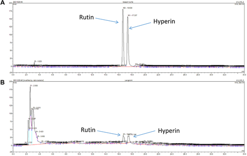
Figure 2 WSS ameliorated pruritus by inhibiting MrgprB2 receptor on mast cell. (A) The skin lesions in mice induced by oxazolone. (B) The WSS could significantly reduce the scratching behavior of ACD model in C57BL/6J mice. (C) The WSS could slightly reduce scratching behavior in ACD model of Sash mice. (D) The WSS could slightly reduce scratching behavior in ACD model of MrgprB2−/− mice (*p < 0.05, **p < 0.01, ***p < 0.001, one-way ANOVA analysis followed by Dunnett’s multiple comparisons test). (E) WSS and ketotifen (10 mg/kg, ig) could reduce the scratching behavior of C57BL/6J mice induced by compound 48/80 (**p < 0.01, ****p < 0.0001, one-way ANOVA analysis followed by Dunnett’s multiple comparisons test). (F and G) WSS had no synergistic effect on the scratching behavior in Sash or MrgprB2−/− mice induced by compound 48/80.
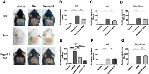
Figure 3 The infiltration of mast cell was inhibited by WSS. (A) Toluidine blue staining of different groups of mice skin tissue of ACD. (B) The number of mast cell was significantly decreased in Oxazolone + WSS group. (C) WSS did not affect the number of mast cells in MrgprB2−/− mice. (D) Toluidine blue staining of different groups of mice skin tissue induced by compound 48/80. (E) The number of mast cell was significantly decreased in C48/80 + WSS group, (F) WSS did not affect the number of mast cells in MrgprB2−/− mice. (*p < 0.05, **p < 0.01, ***p < 0.001, one-way ANOVA analysis followed by Dunnett’s multiple comparisons test), scale bar: 100 μm.
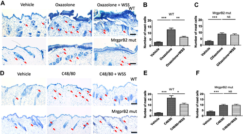
Figure 4 Effect of WSS on the calcium influx caused by compound 48/80 in mouse peritoneal mast cells isolated from C57BL/6J or MrgprB2−/− mice. (A and D) Representative trace for changes in [Ca2+]i induced by compound 48/80 (5 μg/mL), as assayed by Fluo-4 imaging in different extracellular solution. (B and E) The representative traces showing changes in [Ca2+]i as measured by Fluo-4 imaging for C57BL/6J or MrgprB2−/− mice peritoneal mast cells. (C) Compared with the normal external solution, calcium influx induced by compound 48/80 in the solution contain WSS was significantly reduced. (**p < 0.01, 1 mg/mL WSS vs normal, ****p < 0.0001, 3 mg/mL WSS vs normal, one-way ANOVA analysis followed by Dunnett’s multiple comparisons test). (F) WSS had no effect on calcium influx induced by compound 48/80 in mast cells isolated from MrgprB2−/− mice. Scale bar =10 µm.
![Figure 4 Effect of WSS on the calcium influx caused by compound 48/80 in mouse peritoneal mast cells isolated from C57BL/6J or MrgprB2−/− mice. (A and D) Representative trace for changes in [Ca2+]i induced by compound 48/80 (5 μg/mL), as assayed by Fluo-4 imaging in different extracellular solution. (B and E) The representative traces showing changes in [Ca2+]i as measured by Fluo-4 imaging for C57BL/6J or MrgprB2−/− mice peritoneal mast cells. (C) Compared with the normal external solution, calcium influx induced by compound 48/80 in the solution contain WSS was significantly reduced. (**p < 0.01, 1 mg/mL WSS vs normal, ****p < 0.0001, 3 mg/mL WSS vs normal, one-way ANOVA analysis followed by Dunnett’s multiple comparisons test). (F) WSS had no effect on calcium influx induced by compound 48/80 in mast cells isolated from MrgprB2−/− mice. Scale bar =10 µm.](/cms/asset/05eedab5-15a3-4173-aeec-3c85993a0659/djir_a_12199792_f0004_c.jpg)
Figure 5 The effect of WSS on the voltage dependent currents induced by compound 48/80. (A and B) Under the normal extracellular solution and the extracellular solution containing WSS (D and E), typical whole-cell membrane current recorded from a mast cell, which clamped at a holding potential of −20 mV, and 100 ms voltage commands were applied from −130 to +130 mV in 10 mV step. (C) The voltage dependent currents induced by compound 48/80 for mouse peritoneal mast cells in the standard extracellular solution (**p < 0.01, ***p < 0.001, paired Student’s t-test). (F) The voltage dependent currents induced by compound 48/80 for mouse peritoneal mast cells in the extracellular solution contain WSS (*p < 0.05, paired Student’s t-test). (G) The current amplitude in different external solutions (*p < 0.05, **p < 0.01, unpaired Student’s t-test). (H and I) The voltage dependent currents for MrgprB2−/− mast cells in different external solutions (paired Student’s t-test, NS: no significant).
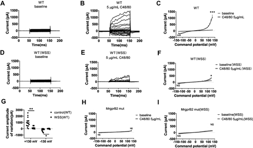
Figure 6 WSS could inhibit the calcium flux induced by compound 48/80 in MrgprB2/X2 over expressing HEK293 cells. (A)The representative traces showing changes in [Ca2+]i in MrgprB2 over-expressing HEK293 cells. (B–D) 300 μg/mL WSS, 1mg/mL WSS, 3 mg/mL WSS inhibited the calcium flux in MrgprB2 over-expressing HEK293 cells induced by compound 48/80. (E) The representative traces showing changes in [Ca2+]i in MrgprX2 over-expressing HEK293 cells. (F–H) 300 μg/mL WSS, 1 mg/mL WSS, 3 mg/mL WSS inhibited the calcium flux in MrgprB2 over-expressing HEK293 cells induced by compound 48/80. (****p < 0.0001, paired Student’s t-test).
![Figure 6 WSS could inhibit the calcium flux induced by compound 48/80 in MrgprB2/X2 over expressing HEK293 cells. (A)The representative traces showing changes in [Ca2+]i in MrgprB2 over-expressing HEK293 cells. (B–D) 300 μg/mL WSS, 1mg/mL WSS, 3 mg/mL WSS inhibited the calcium flux in MrgprB2 over-expressing HEK293 cells induced by compound 48/80. (E) The representative traces showing changes in [Ca2+]i in MrgprX2 over-expressing HEK293 cells. (F–H) 300 μg/mL WSS, 1 mg/mL WSS, 3 mg/mL WSS inhibited the calcium flux in MrgprB2 over-expressing HEK293 cells induced by compound 48/80. (****p < 0.0001, paired Student’s t-test).](/cms/asset/6f28de8c-de6d-4d3b-a6d7-8a4f89f77be1/djir_a_12199792_f0006_c.jpg)