Figures & data
Figure 1 FGF15 treatment supports the survival of septic mice by mitigating hepatic inflammation and liver cell apoptosis. Following the CLP procedure, the mice were randomized and intravenously injected beginning at 2 hours post the procedure every 12 h for three days with saline as the CLP group or FGF15 as the FGF15 group. A sham control group of mice received the sham procedure and saline injection. (A) The survival of different groups of mice. (B) The histopathological changes in the liver of different groups of mice. (C) The percentages of liver cell apoptosis in different groups of mice. Data are representative images or expressed as the mean ± SD of each group (n=9 per group) from three separate experiments.
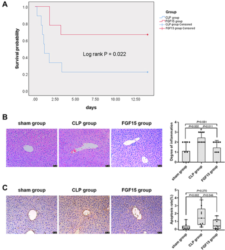
Figure 2 FGF15 treatment mitigates hepatic Treg responses and reduces the levels of serum cytokines in septic mice at 3 days post CLP. (A) Flow cytometric analysis of the frequency of hepatic CD4+CD25+FOXP3+ Tregs in total CD4+ T cells in different groups of mice. (B) Western blot analysis of the relative levels of IL-2, FOXP3 and TGF-β expression in the liver of mice. (C) ELISA analysis of the levels of serum cytokines in different groups of mice. Data are representative images or expressed as the mean ± SD of each group (n=9 per group) from three separate experiments.
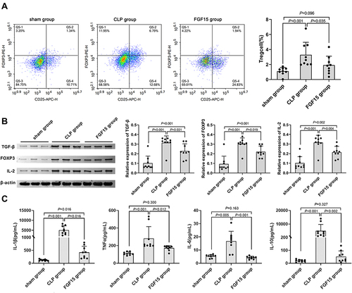
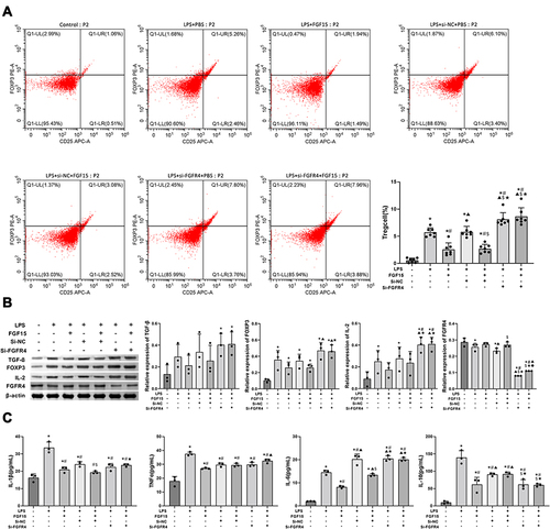
Figure 3 FGF15 treatment mitigates the LPS-enhanced Treg responses in a transwell co-culture system. Liver NCTC 1469 cells were transfected with control or FGFR4-specific siRNA and co-cultured with the isolated liver CD4+ T cells from naïve mice in transwell plates. The cells were stimulated with LPS for 24 h and treated with vehicle saline or FGF15 for 48 h. (A) Flow cytometry analysis of the frequency of CD4+CD25+FOXP3+ Tregs in total CD4+ T cells. (B) Western blot analysis of the relative levels of FGFR4, TGF-β and IL-2 expression in different groups of cells. (C) ELISA analysis of the levels of IL-1β, TNFα, IL-6 and IL-10 in the supernatants of cultured cells. Data are representative images or expressed as the mean ± SD of each group of cells from three separate experiments. *P < 0.05 vs the control group; #P < 0.05 vs the LPS group; ▲P < 0.05 vs the FGF15 group; $P < 0.05 vs the LPS+Si-NC group; ★P < 0.05 vs the LPS+Si-NC+FGF15 group; ◆P < 0.05 vs the LPS+Si-FGFR4 group.