Figures & data
Figure 1 Chest radiograph on admission.
Notes: Large consolidations and multiple lobular shadows were seen in the upper and middle fields of the right lung. The left lung was operated and collapsed due to old tuberculosis.
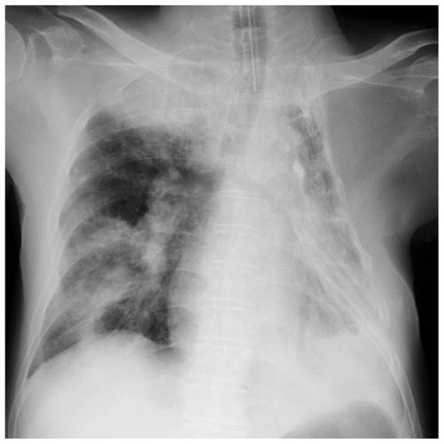
Figure 2 Formation of NETs in the sputum of this patient. (A) 4′,6-diamidino-2-phenylindole staining at high magnification (1000×). (B) Immunohistochemistry against anti-human histone-H3 mouse monoclonal antibody. (C) Immunohistochemistry against anti-human neutrophil elastase rabbit polyclonal antibody. (D) Merged photograph of (A–C). These pictures indicate that the fibrous constituents are NETs. (E) Gram-staining of endotracheal-aspirated sputum at low magnification (1000×).
Note: Arrow indicates numerous fibrous constituents.
Abbreviation: NETs, neutrophil extracellular traps.
Abbreviation: NETs, neutrophil extracellular traps.
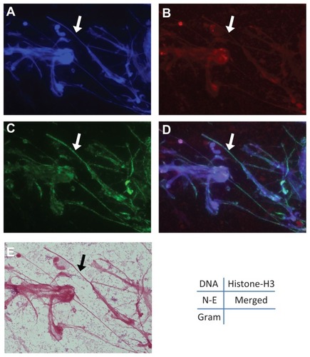
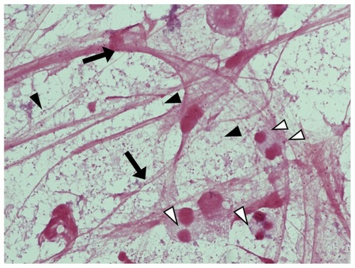
Figure 3 Gram-staining of endotracheal-aspirated sputum.
Notes: A large number of Gram-negative coccobacilli (black arrowheads), including a proportion phagocytosed by neutrophils (white arrowheads) and numerous fibrous NETs (arrows) are observed at higher magnification (1000×).
Abbreviation: NETs, neutrophil extracellular traps.
Abbreviation: NETs, neutrophil extracellular traps.