Figures & data
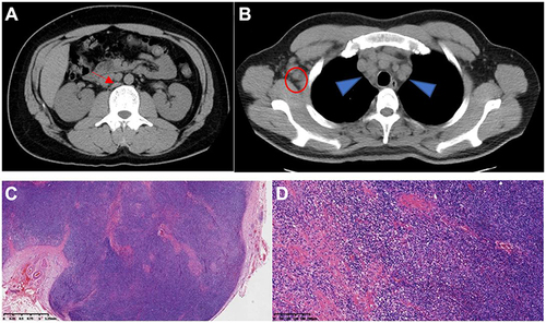
Figure 1 Computed tomography images. (A) swollen lymph nodes in the retroperitoneum (red arrow). (B) Swollen lymph node in right armpit (red circle). Swollen lymph nodes in the mediastinum (blue triangle). (C) Magnification ×20. Lymphoid follicular hyperplasia. (D) Magnification ×100 high endothelial venules with angiogenesis, infiltration of plasma cells and eosinophils, and localized fibrosis.