Figures & data

© 2023 Zhang et al. This work is published by Dove Medical Press Limited, and licensed under Creative Commons Attribution - Non Commerical (unported, v3.0) License. Non-commercial uses of the work are permitted without any further permission from Dove Medical Press Limited, provided the work is properly attributed.
Figure 1 Molecular determinants of the differentiation and polarization of macrophages, and differentiation and maturation of osteoclasts. Macrophages and osteoclasts are the result of competing differentiations of myeloid progenitor. Under the stimulation of M-CSF, myeloid progenitor differentiates into macrophages. Under the dual stimulation of M-CSF and RANKL, myeloid progenitors differentiate into osteoclasts. Under the regulation of multiple factors such as PPARγ, ERRα, PGC-1β, NDUSF4 and maternal VLDLR, the macrophages differentiate into osteoclasts. Different stimuli activate the polarization of M1 and M2 macrophages, which lead to the release of pro-and anti-inflammatory cytokines respectively (“Created with BioRender.com”).
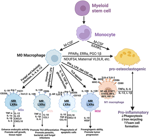
Figure 2 Association between osteogenesis and macrophage polarization in the development of GIONFH. The M0, M1 and M2 phenotypes are all beneficial for osteogenesis by activating the OSM signaling pathway, and the M1-type macrophages had the greatest effect on bone formation. BMSCs secrete CCL-2 and CCL-4, the main chemoattractants for monocytes and macrophages. Meanwhile, Bone macrophages also interact with osteoblasts, which derive from BMSCs (“Created with BioRender.com”).
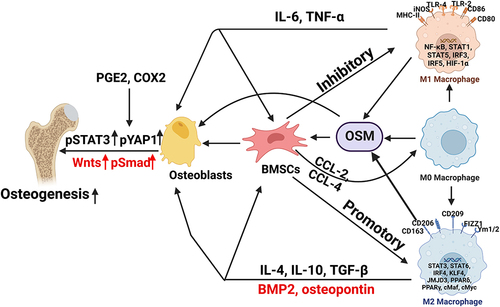
Figure 3 Evaluation of immune cells infiltration in GIONFH. (A) landscape of immune cells infiltration in bone tissue of normal control or GIONFH; (B) correlation analysis among 22 immune cells in necrotic bone samples; (C) violin plot showing the differences on infiltration abundance of 22 immune cells in bone tissue of normal control (cyan) and GIONFH (red).

Figure 4 Activation of M1 polarized macrophage in the development of GIONFH. Damage-associated molecular patterns in the necrotic tissue binds to the TLR4, and subsequently activates Myd88-extracellular regulated kinase 1/2 (ERK1/2), Ik kinase (IKK) α-transcription factors nuclear factor-κB (NF-κB) pathway, which leads to the production of Th1 cytokines (IFN-γ and TNF-α) and HIF-1. The activation of the JAK1/2-STAT1 pathway by IFN-γ and subsequent phosphorylation of STAT1 have also been reported to correlate with the M1 macrophage polarization in GIONFH (“Created with BioRender.com”).
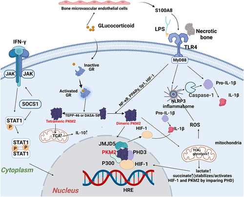
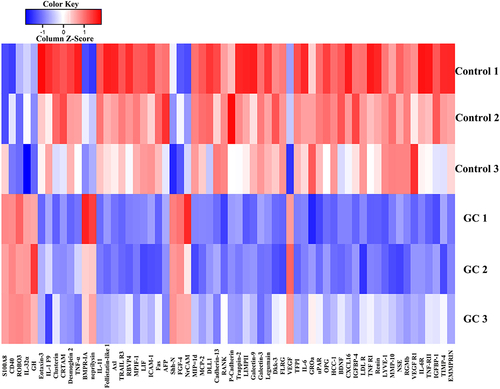
Figure 5 The heatmap about differentially expressed proteins between extracellular vesicles released by bone marrow endothelial cells (BMECs) in the glucocorticoid-induced group and the Control group.