
Figures & data
Figure 1 The effect of macrophages on VSMC calcification. VSMCs were cocultured with the 3 macrophage subtypes in calcification-inducing medium (ANOVA, ****Stands for p<0.0001). (a) Alizarin red staining revealed calcification in the cocultures of different macrophage subtypes and VSMCs (the location of calcification nodules was marked with black arrows). (b) Cetylpyridinium chloride assays were used to quantify calcification levels in the cocultures. M1 macrophages significantly induced calcification in VSMCs.
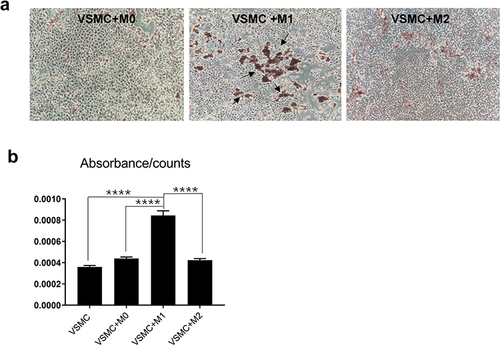
Figure 2 The effect of macrophages on calcification in VSMCs through indirect contact. VSMCs were cultured with M0, M1 or M2 macrophages in Transwells without cell contact in calcification-inducing medium (ANOVA, *Stands for p <0.05, **Stands for p<0.01, and ****Stands for p <0.0001). (a) Alizarin red staining of VSMCs cocultured with different macrophage subtypes (the location of calcification nodules was marked with black arrows). (b) Cetylpyridinium chloride assays showed calcification in the culture medium of the cocultures. (c) The CA1, CA2, Runx2, ALP and BMP mRNA levels in VSMCs were determined using real-time PCR. (d) CA1 and CA2 expression in VSMCs was determined using Western blotting. (e) The levels of IL-1β, IL-6 and TNF-α in the culture medium of VSMCs that were cocultured with different macrophage subtypes were examined using flow cytometry. M1 macrophages could stimulate calcification and elevate CA1 and CA2 expression in VSMCs by indirect contact, while M0 and M1 macrophages, especially M1 macrophages, increased IL-1β, IL-6 and TNF-α levels in the cocultures.
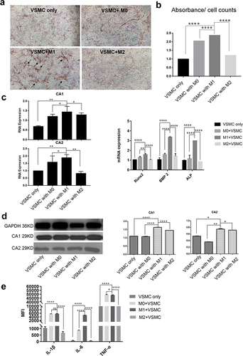
Figure 3 The effect of macrophage supernatants on VSMC calcification. VSMCs were separately cultured with the supernatant of M0, M1 or M2 macrophages and calcification-inducing medium (1:1 proportions) (ANOVA, *Stands for p<0.05, **Stands for p<0.01, ***Stands for p <0.001, and ****Stands for p <0.0001). (a) Alizarin red staining of VSMCs cocultured with supernatants from the different macrophages (the location of calcification nodules was marked with black arrows). (b) Cetylpyridinium chloride assays quantitated calcification levels in VSMCs cocultured with supernatants from the different macrophage subtypes. (c) The mRNA expression of calcification-related genes, including BMP2, ALP and Runx2, in VSMCs was examined by real-time PCR. (d) The mRNA expression of CA1 and CA2 in VSMCs was examined by real-time PCR. (e) CA1 and CA2 protein expression in VSMCs was examined by Western blot analysis. (f) The levels of IL-1β, IL-6 and TNF-α in the culture medium of VSMCs that were cultured with macrophage supernatant were examined by flow cytometry. M1 culture supernatant stimulated calcification, increased CA1 and CA2 expression in VSMCs, and elevated IL-1β, IL-6 and TNF-α levels in the cocultures.
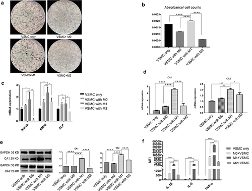
Figure 4 The effect of anti-CA1 and anti-CA2 siRNA on VSMC calcification. VSMCs were transfected with anti-CA1 and anti-CA2 siRNA and cultured in M1 macrophage supernatant and calcification-inducing medium (1:1) (ANOVA, *Stands for p<0.05, **Stands for p<0.01, ***Stands for p <0.001, and ****Stands for p <0.0001). (a) Real-time PCR indicated the successful inhibition of target gene expression in VSMCs by anti-CA1 and anti-CA2 siRNAs. (b) AR-S staining showed few calcified nodules in VSMCs that were transfected with anti-CA1, and M1 culture supernatants partially restored calcification levels (the location of calcification nodules was marked with black arrows). Quantification of calcification with a cetylpyridine chloride assay verified this result. (c) AR-S staining showed few calcified nodules in VSMCs that were transfected with anti-CA2 siRNA, and M1 culture supernatants partially restored calcification levels (the location of calcification nodules was marked with black arrows). Quantification of calcification with a cetylpyridine chloride assay verified this result. CA1 and CA2 expression plays an essential role in the calcification of VSMCs.
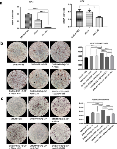
Figure 5 The effect of anti-CA1 and anti-CA2 siRNA on CA1 and CA2 expression in VSMCs and proinflammatory cytokine production (ANOVA, *Stands for p<0.05, **Stands for p<0.01, ***Stands for p <0.001, and ****Stands for p <0.0001). (a) CA1 expression in VSMCs was examined by real-time PCR. (b) CA1 expression in VSMCs was examined by Western blotting. (c) CA2 expression in VSMCs was examined by real-time PCR. (d) CA2 expression in VSMCs was examined by Western blotting. Anti-CA1 or anti-CA2 siRNA treatment decreased CA1 and CA2 expression in VSMCs, respectively, and M1 supernatant alleviated the suppressive effects of anti-CA1 and anti-CA2 siRNA on the target genes. (e) The levels of IL-6, IL-1β and TNF-α in the culture medium of VSMCs after transfection with anti-CA1. (f) The levels of IL-6, IL-1β and TNF-α in the culture medium of VSMCs after transfection with anti-CA2. Anti-CA1 and anti-CA2 siRNA transfection reduced IL-6, IL-1β and TNF-α levels in the culture, and M1 supernatant alleviated the suppressive effects of anti-CA1 and anti-CA2 siRNA on proinflammatory cytokine production.
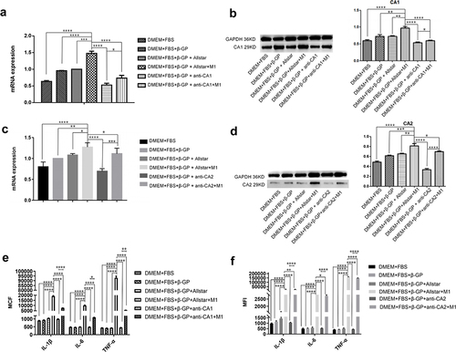
Figure 6 The effect of TNF-α on calcification and CA1 and CA2 expression in VSMCs (ANOVA, *Stands for p<0.05, **Stands for p<0.01, ***Stands for p <0.001, and ****Stands for p <0.0001). (a) The expression of CA1, CA2 and three calcification-associated genes in VSMCs treated with different concentrations of TNF-α was examined by real-time PCR. (b) Alizarin red staining of VSMCs treated with TNF-α (10 ng/mL). (c) Quantification of calcification in VSMCs treated with TNF-α (10 ng/mL) (the location of calcification nodules was marked with black arrows). (d) CA1 and CA2 expression in VSMCs was examined by Western blot analysis. TNF-α could induce calcification and CA1 and CA2 expression in VSMCs without β-GP induction.
