Figures & data
Figure 1 The inclusive process of the participants.
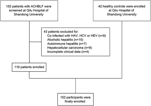
Figure 2 The expression of lncRNA GAS5 in serum exosomes was elevated in patients with ACHBLF. (a) The representative image of serum exosomes analyzed by TEM. (b) Exosomal markers CD81 and Hsp70 were detected using Western blot. (c) The expression level of serum exosomal lncRNA GAS5 in patients with ACHBLF and HCs.
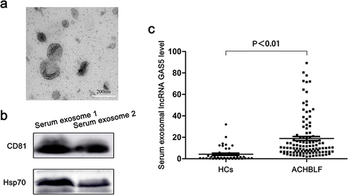
Table 1 Baseline Characteristics of the Enrolled Participants
Table 2 Relationship Between lncRNA GAS5 Expression and Clinicopathological Characteristics in ACHBLF
Figure 3 Over-expressed serum exosomal lncRNA GAS5 was associated with adverse clinicopathological characteristics and poor prognosis in ACHBLF. (a–d) Statistical correlation analysis between the expression level of serum exosomal lncRNA GAS5 and TBIL (a), DBIL (b), PT-INR (c) and PTA (d). (e) Kaplan–Meier curves for 3-month mortality of patients with ACHBLF grouped according to relative serum exosomal lncRNA GAS5 expression. (f and g) ROC curves of serum exosomal lncRNA GAS5 level (f) and MELD score (g) in predicting 3-month mortality of ACHBLF.
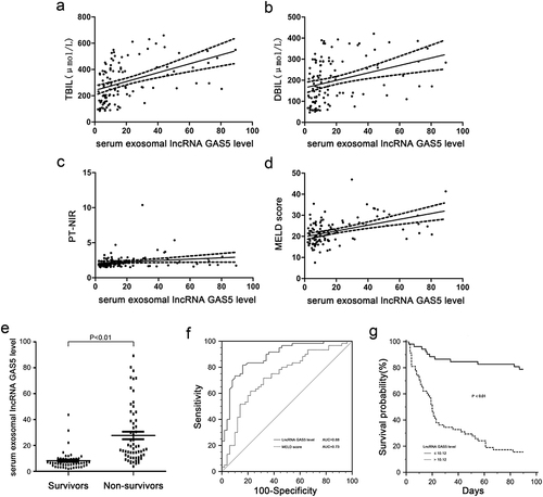
Table 3 Univariate and Multivariate Analysis of Factors Associated with 3-Month Mortality of Patients with ACLF
Figure 4 LncRNA GAS5 inhibited hepatocytes proliferation and increased hepatocytes apoptosis. (a) Relative expression of lncRNA GAS5 after MIHA cells transfected with pIRES2-NC, pIRES2-GAS5. (b) Relative expression of lncRNA GAS5 after MIHA cells transfected with si-NC or si-GAS5. (c) Cell viability of MIHA cells transfected with pIRES2-NC, pIRES2-GAS5, si-NC or si-GAS5. (c) Cell apoptosis of MIHA cells transfected with pIRES2-NC, pIRES2-GAS5, si-NC or si-GAS5. (d) Cell apoptosis of MIHA cells transfected with pIRES2-NC, pIRES2-GAS5, si-NC or si-GAS5. *P < 0.05, **P < 0.01, ***P < 0.001.
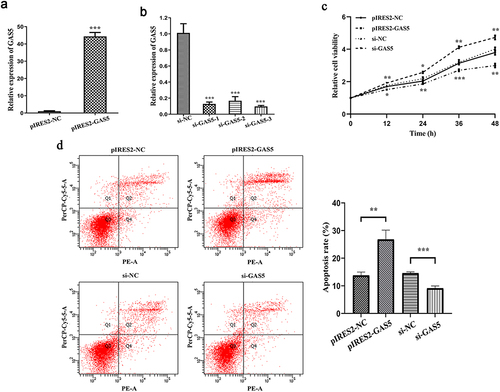
Figure 5 Exosomes-mediated lncRNA GAS5 transfer promoted hepatocytes injury. (a) The representative images of exosomes isolated from conditioned medium of MIHA cells. (b) The size distribution of isolated exosomes. (c) Exosomal markers CD81 and Hsp70 were detected by Western blot. (d) The representative images of MIHA cells cultured in the absence (control) or presence of PKH67-labeled exosomes. (e) Relative expression of lncRNA GAS5 in exosomes isolated from conditioned medium of MIHA cells overexpressing lncRNA GAS5 (GAS5-exo) or control cells (NC-exo). (f) Relative expression of lncRNA GAS5 in MIHA cells incubated with GAS5-exo or NC-exo. (g) Cell viability of MIHA cells incubated with GAS5-exo or NC-exo. (h) Cell apoptosis of MIHA cells incubated with GAS5-exo or NC-exo. *P < 0.05, **P < 0.01, ***P < 0.001.
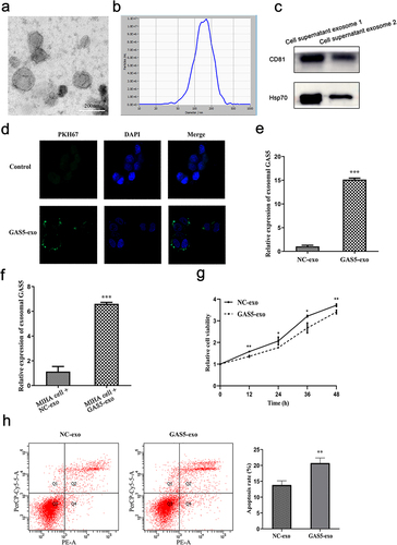
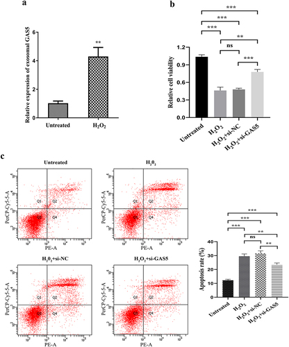
Figure 6 Knocked down of lncRNA GAS5 weakened H2O2-induced hepatocytes injury. (a) Relative expression of lncRNA GAS5 in H2O2 treated MIHA cells or control cells. (b) Cell viability of H2O2 treated MIHA cells or control cells with or without lncRNA GAS5 knockdown. (c) Cell apoptosis of H2O2 treated MIHA cells or control cells with or without lncRNA GAS5 knockdown. **P < 0.01, ***P < 0.001, ns: not significant.