Figures & data
Figure 1 (A) At 24 weeks pregnant. Suprasternal view of aortic arch shows the wall continuity from the aortic arch to the descending aorta is intact. The green box represents the Color Doppler sampling position. Blue blood flow signal shows no evident anomaly in blood flow of the aortic arch and descending segment. (B) 15 days after birth. There is stenosis in the aortic arch starting from the opening of the LCCA, with the distal end being a blind end. There is stenosis at the opening of the LSCA. White arrow represents the distal end of the transverse aortic arch was a blind end.
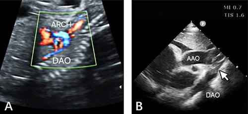
Figure 2 (A) CT angiography. White arrow represents the lumen of the aortic arch’s descending transition segment is obstructed, with an affected length of approximately 0.45 cm. (B) Vascular 3D reconstruction techniques. White arrow represents the atretic aortic arch. No finer bundles were detected in the atretic areas.
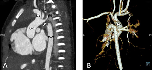
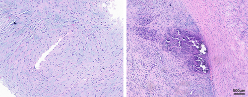
Figure 3 Arterial wall tissue had extensive mucoid degeneration with poor muscular structure and calcification and granulation tissue in some areas.