Figures & data
Figure 1 (a) Study flow chart. (b) Patients categorization chart.
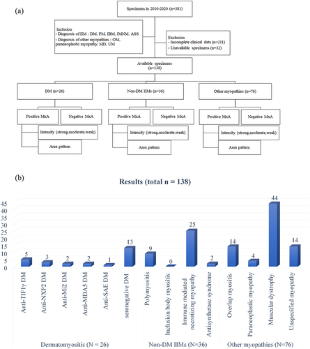
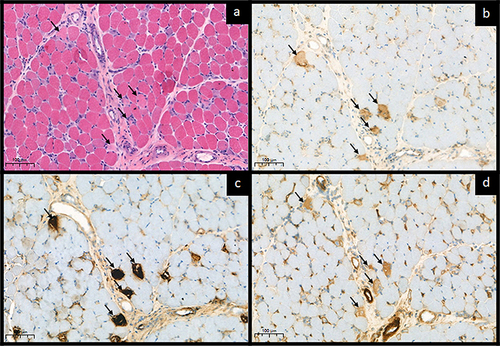
Figure 2 Representative immunohistochemical staining of MxA in muscle biopsies. Positive expression was defined by unequivocal sarcoplasmic staining in only intact fiber, not a regenerating or degenerated/necrotic fiber. (a) MxA negativity. (b) Moderate MxA positivity and perifascicular area pattern. (c) Strong MxA positivity and perifascicular area pattern (b and c in dermatomyositis patients). (d) Weak MxA positivity and non-perifascicular area pattern, positive fibers labeled with black arrow (in overlap myositis patient).
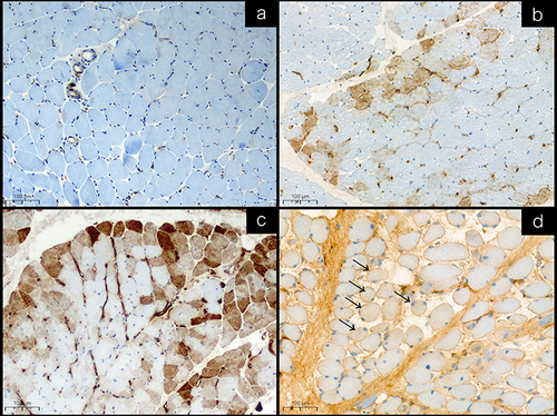
Table 1 Demographic Data, Diagnosis and Myositis-Specific Autoantibodies of 138 Patients
Table 2 The Details of MxA Staining on Muscle Biopsy Specimens of Patients with Different Diagnosis
Figure 3 Necrotic fibers features on MxA and other IHCs. Sarcoplasmic MxA expression interpretation should be rigorous. (a) Five atrophic/shrinkage fibers with pale cytoplasmic stain were identified on H&E, 20X (black arrows). (b) The fibers revealed equivocal sarcoplasmic expression on MxA (black arrows). (c) The corresponding fibers revealed strong/clumping stained on MAC, 20X (black arrows). (d) Strong sarcoplasmic utrophin upregulation, 20X (black arrows). All stains represented that they were degenerated/necrotic fibers.