Figures & data
Figure 1 Picture of the duodenal bulb showing an ulcer with a central naked thrombus head and active bleeding on the surface.
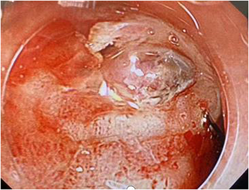
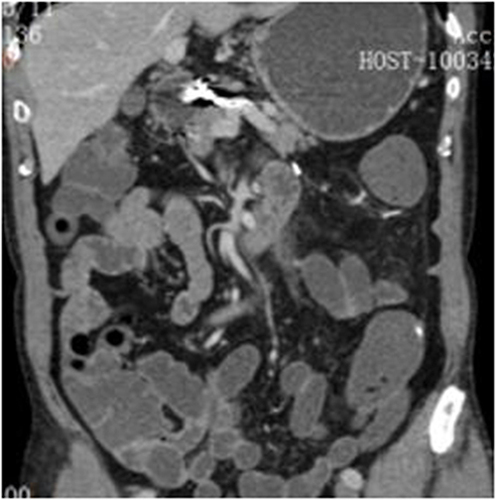
Figure 2 Initial computed tomography image showing no abnormalities in the abdomen.
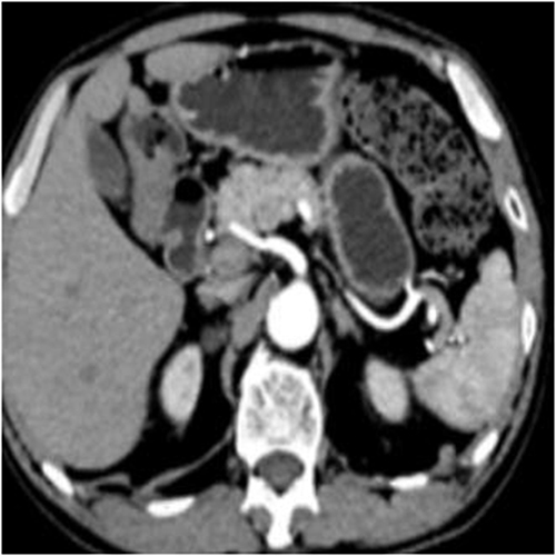
Figure 3 Angiography showing the presence of a distal tumor dilatation at the proximal branch of the common hepatic artery.
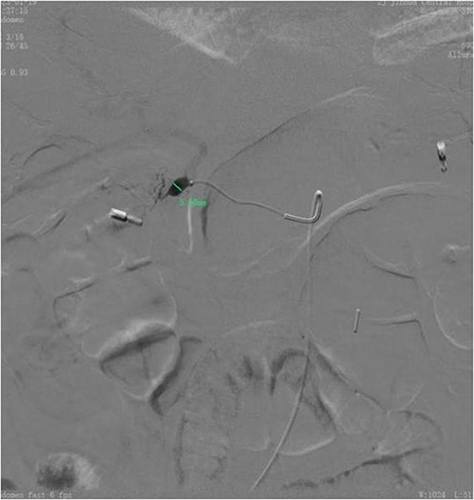
Figure 4 Image showing the embolization of the common hepatic artery.
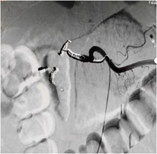
Figure 5 Disease diagnosis and treatment procedure.
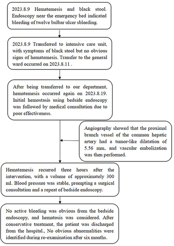
Figure 6 A follow-up gastroscopic examination after six months showed scarring changes in the duodenal bulbar ulcer.
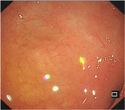
Figure 7 Reexamination based on an abdominal computed tomography scan showed postoperative changes in the proximal branch aneurysms of the common hepatic artery.