
Figures & data
Figure 1 Representative photomicrographs of skin biopsies submitted for Southwestern histochemistry for detection of activated NFκB (original magnification 200×).
Notes: Arrows demonstrate purple nuclear staining. Detection was quantified by an NFκB activation index, which values ranged from zero to four. (A) NFκB activation index of 0: no nuclear staining (negative control); (B) NFκB activation index of 1: up to 10% nuclear staining in sparse lymphocytes from a tuberculoid lesion; (C) NFκB activation index of 2: 11%–25% nuclear staining in cells from a superficial lymphohistiocytic infiltrate and fibroblasts (arrows) from a borderline tuberculoid lesion; (D) NFκB activation index of 3: 26%–50% nuclear staining in cells from a perivascular and perineural inflammatory infiltrate, involving the superficial and mid dermis. There was staining of histiocytes, lymphocytes, fibroblasts, and vascular endothelium (arrow tip) from a lepromatous lesion; (E) NFκB activation index of 4: >50% nuclear staining in cells from a dense perineural and perivascular inflammatory infiltrate. There was staining of fibroblasts, histiocytes, and lymphocytes from a borderline lesion.
Abbreviation: NFκB, nuclear factor kappa B.
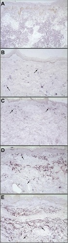
Table 1 Data from 38 leprosy patients evaluated for activation of NFκB
Table 2 Logistic regression analysis of activated NFκB according to leprosy classification, adjusted by age group and sex
Table 3 Distribution of NFκB activation indexes by leprosy classification and WHO classification