Figures & data
Figure 1 HLA-DR-ir microglia morphology and semiquantitative rating. Representative high magnification images of HLA-DR-positive resident (A) and activated (B) microglia. Characteristic images of HLA-DR-ir nigral sections showing a score of 1 (C) and 4 (D) on a rating scale of 0–4 defining the presence of activated microglia. Scale bar: (A and B) =10 μm; (C and D) =50 μm.
Abbreviations: HLA, human leukocyte antigen; ir, immunoreactivity.
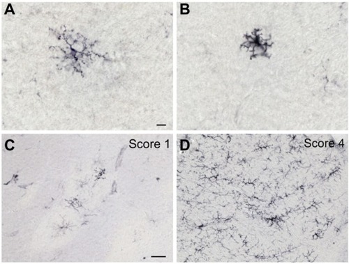
Figure 2 TH-ir nigral cell counts and volumes are variable in 6-OHDA-treated monkeys. Microphotographs of TH immunostained striatum at the level of the anterior commissure of control (A) and 6-OHDA-treated (B) monkeys, and in the left substantia nigra of a control (C, c), a 6-OHDA-treated monkey without reaction to toxin (D, d), and a 6-OHDA-treated monkey with a mild reaction to toxin (E, e). Stereological analysis of averaged TH-ir nigral cell counts and volumes show no difference between groups (F). TH-ir nigral cell counts for individual animals show no significant difference in the control group, but significant differences were found in 6-OHDA-treated animals, with r01098, rh2316, and rh2318 showing a smaller number of nigral cells than r04094 (G). TH-ir nigral cell volumes for individual animals show no significant difference in the control group, but significant differences were found in 6-OHDA-treated animals, with a lower nigral volume found in r01097 and r01098 compared with r04094 and decreased volumes in rh2316 and rh2318 compared with all other 6-OHDA-treated animals (H). *P<0.05, **P<0.01, ***P<0.001. Scale bar: (A and B) =10 mm, (C–E) =500 μm; insets =10 μm.
Abbreviations: 6-OHDA, 6-hydroxydopamine; TH, tyrosine hydroxylase; ir, immunoreactivity.
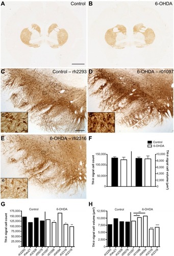
Figure 3 Distribution of α-synuclein-ir varies in the substantia nigra. Representative images of α-synuclein immunostaining in nigral cell bodies (A, a) or fibers (B, b). Scale bar: (A and B) =500 μm; (a and b) =50 μm.
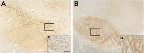
Figure 4 Increased expression of HLA-DR in blood vessels of 6-OHDA-treated monkeys. Microphotographs of HLA-DR immunostained striatum at the level of the anterior commissure of a control monkey (A) and a 6-OHDA-treated monkey (B). Mild HLA-DR expression was found in the cerebrovasculature of a control animal in the caudate (C) and hippocampus (E, e), while HLA-DR-ir was upregulated in 6-OHDA animals in the caudate (D) and hippocampus (F, f). HLA-DR-ir blood vessel scores were significantly higher in the caudate, septum, and hippocampus of 6-OHDA-treated animals compared with controls (G). Scores for HLA-DR-ir blood vessels negatively correlated with TH-ir nigral cell counts (H). *P<0.05. Scale bar: (A and B) =10 mm, (C–F) =500 μm; insets =100 μm.
Abbreviations: 6-OHDA, 6-hydroxydopamine; BV, blood vessel; HLA, human leukocyte antigen; TH, tyrosine hydroxylase; ir, immunoreactivity.
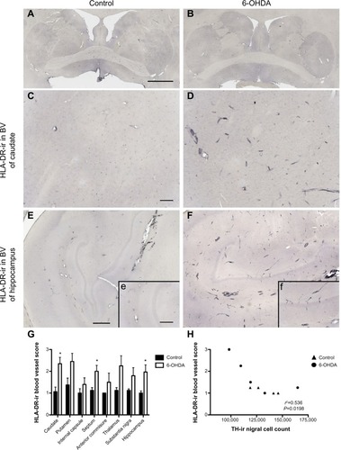
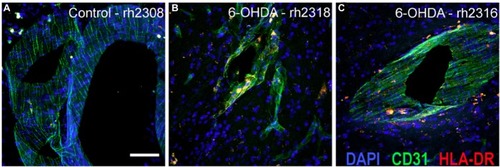
Figure 5 Immunofluorescent staining localizes HLA-DR-ir in CD31+ vasculature. Immunofluorescence labeling demonstrated increased HLA-DR-ir threaded in CD31-positive blood vessels of the ventral putamen in animals that received systemic 6-OHDA (B and C) compared with controls (A). Scale bar =25 μm.
Abbreviations: 6-OHDA, 6-hydroxydopamine; CD31, platelet endothelial cell adhesion molecule; DAPI, 4′,6-diamidino-2-phenylindole; HLA, human leukocyte antigen; ir, immunoreactivity.