Figures & data
Figure 1 Consequences of tissue trauma on heme catabolism, reactive oxygen species, and polyunsaturated fatty acid metabolism.
Notes: Top panel: following tissue trauma, release of free heme can serve as a signal for proinflammatory events.Citation71 The oxidative degradation of heme by heme oxygenase removes free heme, and generates catabolites with antioxidant and anti-inflammatory effects.Citation70–Citation75 Iron released from heme by heme oxygenase catabolism can be stored by ferritin, preventing its participation in redox reactions.Citation70 Middle panel: acute inflammation is associated with an increase in reactive oxygen species, which are regulated by cellular enzymes involved in redox reactions.Citation55,Citation76,Citation77 Reactive oxygen species can have proinflammatory effects, and chronic inflammatory diseases are often associated with oxidative stress.Citation55,Citation76,Citation77,Citation79 In some cases, reactive oxygen species can have anti-inflammatory effects.Citation62,Citation78 lower panel: in response to proinflammatory stimuli, phospholipase A2 mediates release of arachidonic acid from the cell membrane.Citation80,Citation81 an increase in inducible prostaglandin-endoperoxide synthase 2 also occurs.Citation80,Citation82–Citation84 During the early inflammatory response, arachidonic acid is metabolized by prostaglandin synthases and lipoxygenases to generate eicosanoids and leukotrienes,Citation21,Citation24,Citation85–Citation87 which is followed, at later times, by “lipid mediator class switching.” The latter is thought to serve as a switch from the production of proinflammatory lipid mediators to those involved in programmed resolution.Citation10,Citation16,Citation19–Citation22
Abbreviations: GSH, reduced glutathione; GSSG, glutathione disulfide; PGD2, prostaglandin D2; PGE2, prostaglandin E2; PGG2, prostaglandin G2; PGH2, prostaglandin H2; PGI2, prostaglandin I2; PUFA, polyunsaturated fatty acid.
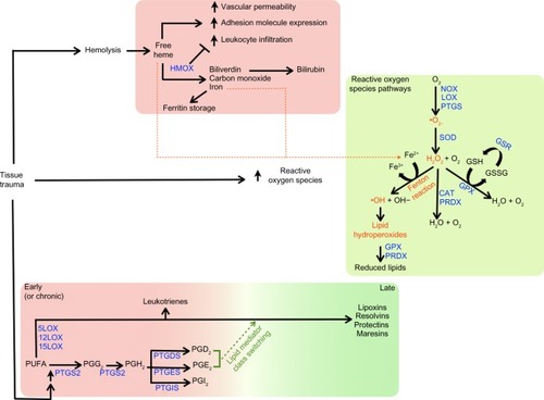
Figure 2 An illustration of the device’s spiral antenna.
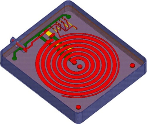
Table 1 List of messenger RNAs evaluated in the study
Table 2 Statistically meaningful changes in mean mRNA levels detected within 4 hours after PEMF treatment relative to untreated control cells
Figure 3 The effect of PEMF treatment on heme oxygenase mRNA levels in HEK, HDF, and HMNC in culture.
Notes: Cells were treated with PEMF for 30 minutes, after which total RNA was isolated and mRNAs quantitated at several time points by qRT-PCR. Cells were harvested at the time points indicated (A) or 2 hours postinitiation of PEMF treatment (B). In (A), the x axis indicates the amount of time passed after the initiation of PEMF treatment, with the end of the 30-minute treatment period indicated by an arrow. In (B), the results of three independent experiments with PEMF are shown. All data is expressed relative to untreated control cell cultures.
Abbreviations: HDF, human dermal fibroblasts; HEK, human epidermal keratinocytes; HMNC, human mononuclear cells; mRNA, messenger RNA; PEMF, pulsed electromagnetic field; qRT-PCR, quantitative reverse-transcription polymerase chain reaction.
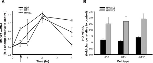
Figure 4 Changes in cellular redox enzyme mRNA levels following PEMF treatment of HDF, HEK, and HMNC.
Notes: Cells were treated with PEMF for 30 minutes, harvested 2 hours after treatment, and relative mRNA levels were determined by qRT-PCR. All data are expressed relative to untreated control cell cultures. n=6.
Abbreviations: HDF, human dermal fibroblasts; HEK, human epidermal keratinocytes; HMNC, human mononuclear cells; mRNA, messenger RNA; PEMF, pulsed electromagnetic field; qRT-PCR, quantitative reverse-transcription polymerase chain reaction.
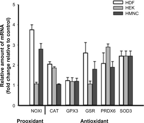
Figure 5 The effect of PEMF on RNA message levels of enzymes involved in lipid mediator synthesis.
Notes: HEK, HDF, and HMNC were treated with PEMF for 30 minutes, after which relative amounts of mRNAs encoding (A) prostaglandin synthases and (B) lipoxygenases were determined using qRT-PCR. Cells were harvested 4 hours (A) or 2 hours (B) after initiation of PEMF treatment. n=6.
Abbreviations: HDF, human dermal fibroblasts; HEK, human epidermal keratinocytes; HMNC, human mononuclear cells; mRNA, messenger RNA; PEMF, pulsed electromagnetic field; qRT-PCR, quantitative reverse-transcription polymerase chain reaction.
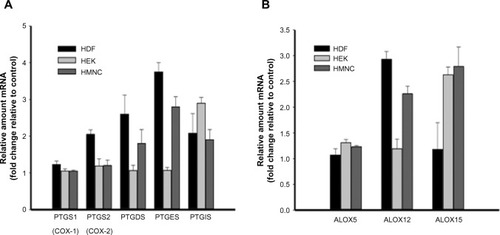
Figure 6 The effect of PEMF treatment on cytokine mRNA levels.
Notes: HEK, HDF, or HMNC were treated with PEMF for 30 minutes. Total RNA was isolated 2 hours after PEMF treatment, and relative amounts of cytokine mRNA were determined using qRT-PCR. n=6–9. All data is expressed relative to untreated control cell cultures.
Abbreviations: HDF, human dermal fibroblasts; HEK, human epidermal keratinocytes; HMNC, human mononuclear cells; mRNA, messenger RNA; PEMF, pulsed electromagnetic field; qRT-PCR, quantitative reverse-transcription polymerase chain reaction.
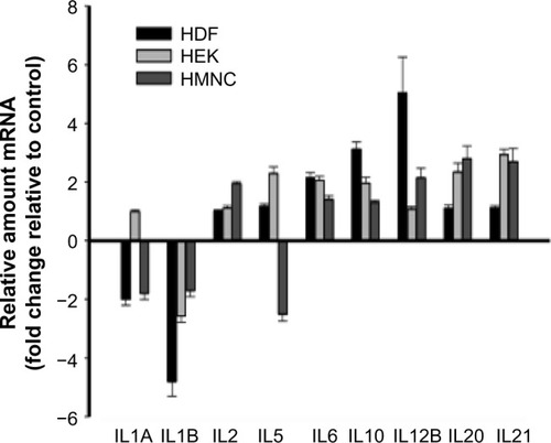
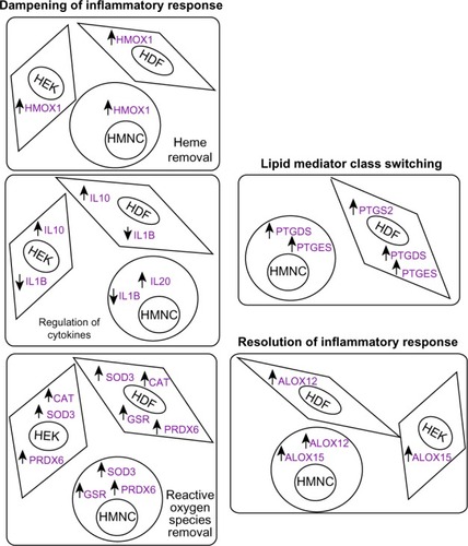
Figure 7 Summary of experimental results from the current study in the context of a possible model.
Notes: In the current study, PEMF treatment was followed by changes in relative amounts of mRNAs encoding factors associated with anti-inflammatory and proresolution effects. An increase or decrease in mRNA levels detected following PEMF treatment, relative to untreated control cells, is indicated by upward- and downward-facing arrows, respectively.
Abbreviations: HDF, human dermal fibroblasts; HEK, human epidermal keratinocytes; HMNC, human mononuclear cells; PEMF, pulsed electromagnetic field; mRNA, messenger RNA.