
Figures & data
Figure 1 Rudolf Virchow (1821–1902).
Note: Photo courtesy of Carl Günther (public domain).Citation105
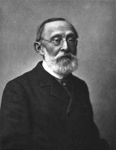
Figure 2 Carl von Rokitansky (1804–1878).
Note: Photo courtesy of Adolf Dauthage (public domain).Citation106
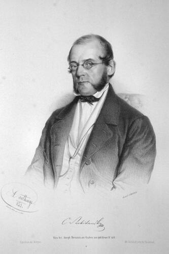
Figure 3 Schematic of atherogenesis.
Notes: The oxidized-LDLs induce the activation of endothelial cells and the expression of various leukocyte adhesion molecules (such as VCAM1) and consequent monocyte adhesion to the endothelium (1,2); subsequent transmigration of monocytes into the intima, where they differentiate into macrophages (3,4); T lymphocytes join macrophages in the intima during plaque evolution (5); macrophages, incorporating modified lipoproteins, become lipid-rich foam cells (6); the inflammatory response stimulates migration and replication of vascular smooth muscle cells, which accumulate in the plaque to form a fibroproliferative lesion (7,8); macrophages in the plaque show abnormal lipid metabolism with a reduction of the cholesterol efflux, (9) which leads to accumulation of apoptotic bodies and necrotic debris, forming a necrotic core (10). ©[designua]/123RF.COM
![Figure 3 Schematic of atherogenesis.Notes: The oxidized-LDLs induce the activation of endothelial cells and the expression of various leukocyte adhesion molecules (such as VCAM1) and consequent monocyte adhesion to the endothelium (1,2); subsequent transmigration of monocytes into the intima, where they differentiate into macrophages (3,4); T lymphocytes join macrophages in the intima during plaque evolution (5); macrophages, incorporating modified lipoproteins, become lipid-rich foam cells (6); the inflammatory response stimulates migration and replication of vascular smooth muscle cells, which accumulate in the plaque to form a fibroproliferative lesion (7,8); macrophages in the plaque show abnormal lipid metabolism with a reduction of the cholesterol efflux, (9) which leads to accumulation of apoptotic bodies and necrotic debris, forming a necrotic core (10). ©[designua]/123RF.COM](/cms/asset/14d878d8-92c5-44e1-8d49-ec582b6e2db0/djmd_a_12169461_f0003_c.jpg)