
Figures & data
Figure 1 The study profile. SCC indicates spinal cord compression; MESCC indicates metastatic epidural spinal cord compression.
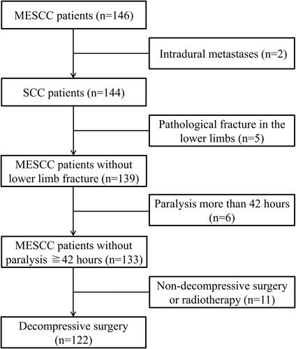
Figure 2 A 47-year-old female diagnosed with lung cancer treated with surgical decompression and pedicle stabilization combined with 125I brachytherapy seeds implantation. (A) Preoperative lateral X-ray showed T6 and T8 vertebral collapse. (B) Preoperative anteroposterior X-ray showed T6 and T8 vertebral collapse. (C) Preoperative lateral CT showed T6 and T8 bone destruction. (D) Preoperative lateral MRI showed T6 and T8 spine metastases. (E) Preoperative transverse CT showed bone destruction at T8. (F) Preoperative transverse MRI showed deformation of the dural sac due to metastatic tumor.
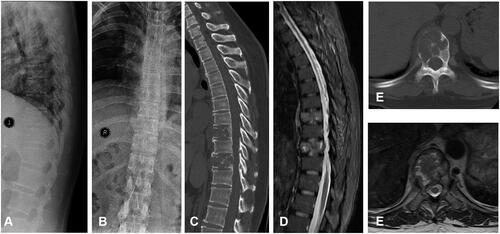
Figure 3 (A) Postoperative lateral X-ray showed 125I brachytherapy seeds implantation and pedicle stabilization with screws. (B) Postoperative anteroposterior X-ray showed 125I brachytherapy seeds implantation and pedicle stabilization with screws. (C) Anterior bone scan at postoperative 10 months. (D) Posterior bone scan at postoperative 19 months. (E) Anterior bone scan at postoperative 19 months. (F) Posterior bone scan at postoperative 19 months. The above bone scan indicated no disease progression at 19 months after surgery.
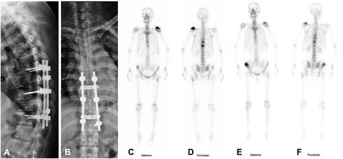
Table 1 Patient’s Characteristics of the Brachytherapy and Irradiation Group
Table 2 The Comparison of Intraoperative Variables and Postoperative Clinical Outcomes Between the Brachytherapy and Irradiation Group
Figure 4 The mean VAS score in a 24-hour period before surgery and at the time of discharge, 1 month, three months, and six months after surgery in both groups. “pre-” indicates pre-operation; “disch-” indicates discharge; “m” indicates months; *Indicates P˂0.05; ***Indicates P˂0.01.
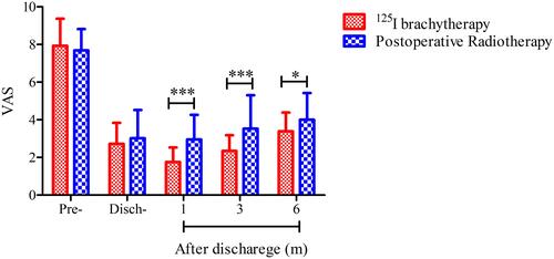
Figure 5 The postoperative overall survival curves for the two groups (P=0.37, Log rank test).
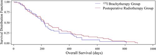
Table 3 Univariate and Multivariate Analyses of Preoperative Factors for Postoperative Overall Survival in MESCC Patients