Figures & data
Box 1 Mayo Clinic Criteria for TTS
Box 2 Heart Failure Association Diagnostic Criteria for TTS
Box 3 International Takotsubo Diagnostic Criteria (InterTAK Diagnostic Criteria)
Table 1 Important Signs in TTS Patients Indicating a Need for Consultation with Specialists
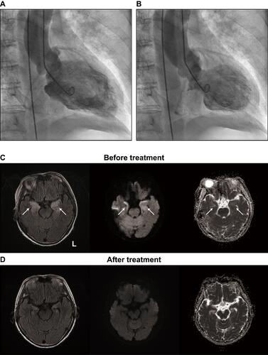
Figure 1 TTS following autoimmune limbic encephalitis. (A and B) Left ventriculographic findings. The diastole (A) and systole (B) images are shown. (C and D) MRI findings. The initial MRI (C) revealed abnormal hyperintense areas (arrows) in the bilateral medial temporal lobes on the fluid-attenuated inversion recovery image (left). Diffusion-weighted imaging (middle) and an apparent diffusion coefficient map (right) both revealed abnormal hyperintensity in these areas. The abnormalities were all resolved in follow-up MRI (D).